
Page 468 - Feline diagnostic imaging
P. 468
480 27 Urinary Disease
(a) (b)
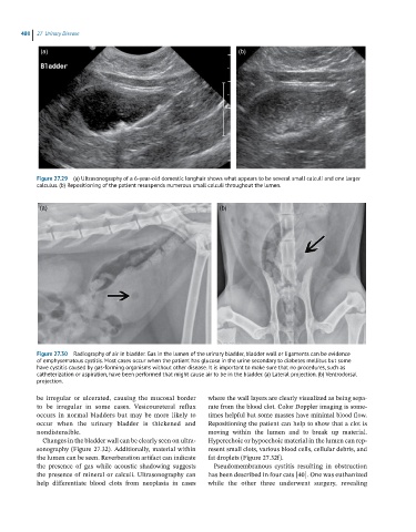
Figure 27.29 (a) Ultrasonography of a 6-year-old domestic longhair shows what appears to be several small calculi and one larger
calculus. (b) Repositioning of the patient resuspends numerous small calculi throughout the lumen.
(a) (b)
Figure 27.30 Radiography of air in bladder. Gas in the lumen of the urinary bladder, bladder wall or ligaments can be evidence
of emphysematous cystitis. Most cases occur when the patient has glucose in the urine secondary to diabetes mellitus but some
have cystitis caused by gas-forming organisms without other disease. It is important to make sure that no procedures, such as
catheterization or aspiration, have been performed that might cause air to be in the bladder. (a) Lateral projection. (b) Ventrodorsal
projection.
be irregular or ulcerated, causing the mucosal border where the wall layers are clearly visualized as being sepa-
to be irregular in some cases. Vesicoureteral reflux rate from the blood clot. Color Doppler imaging is some-
occurs in normal bladders but may be more likely to times helpful but some masses have minimal blood flow.
occur when the urinary bladder is thickened and Repositioning the patient can help to show that a clot is
nondistensible. moving within the lumen and to break up material.
Changes in the bladder wall can be clearly seen on ultra- Hyperechoic or hypoechoic material in the lumen can rep-
sonography (Figure 27.32). Additionally, material within resent small clots, various blood cells, cellular debris, and
the lumen can be seen. Reverberation artifact can indicate fat droplets (Figure 27.32f).
the presence of gas while acoustic shadowing suggests Pseudomembranous cystitis resulting in obstruction
the presence of mineral or calculi. Ultrasonography can has been described in four cats [40]. One was euthanized
help differentiate blood clots from neoplasia in cases while the other three underwent surgery, revealing