
Page 472 - Feline diagnostic imaging
P. 472
484 27 Urinary Disease
(a) (b)
(c)
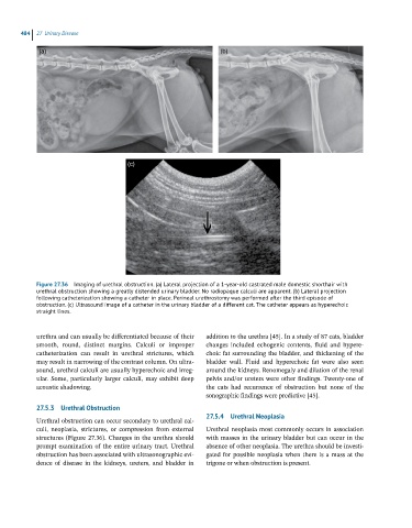
Figure 27.36 Imaging of urethral obstruction. (a) Lateral projection of a 1-year-old castrated male domestic shorthair with
urethral obstruction showing a greatly distended urinary bladder. No radiopaque calculi are apparent. (b) Lateral projection
following catheterization showing a catheter in place. Perineal urethrostomy was performed after the third episode of
obstruction. (c) Ultrasound image of a catheter in the urinary bladder of a different cat. The catheter appears as hyperechoic
straight lines.
urethra and can usually be differentiated because of their addition to the urethra [45]. In a study of 87 cats, bladder
smooth, round, distinct margins. Calculi or improper changes included echogenic contents, fluid and hypere-
catheterization can result in urethral strictures, which choic fat surrounding the bladder, and thickening of the
may result in narrowing of the contrast column. On ultra- bladder wall. Fluid and hyperechoic fat were also seen
sound, urethral calculi are usually hyperechoic and irreg- around the kidneys. Renomegaly and dilation of the renal
ular. Some, particularly larger calculi, may exhibit deep pelvis and/or ureters were other findings. Twenty‐one of
acoustic shadowing. the cats had recurrence of obstruction but none of the
sonographic findings were predictive [45].
27.5.3 Urethral Obstruction
27.5.4 Urethral Neoplasia
Urethral obstruction can occur secondary to urethral cal-
culi, neoplasia, strictures, or compression from external Urethral neoplasia most commonly occurs in association
structures (Figure 27.36). Changes in the urethra should with masses in the urinary bladder but can occur in the
prompt examination of the entire urinary tract. Urethral absence of other neoplasia. The urethra should be investi-
obstruction has been associated with ultrasonographic evi- gated for possible neoplasia when there is a mass at the
dence of disease in the kidneys, ureters, and bladder in trigone or when obstruction is present.