
Page 476 - Feline diagnostic imaging
P. 476
488 28 Reproduction
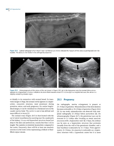
Figure 28.1 Lateral radiograph of an intact 3-year-old female cat. Arrows indicate the margins of the uterus superimposed over the
bladder. The uterus is not visible in the orthogonal projection.
(a) (b)
Figure 28.2 Ultrasonography of the uterus of the cat shown in Figure 28.1. (a) In the transverse scan, the normal feline uterus
appears as a hypoechoic circular or elliptical structure that measures about 0.5–1.0 cm. (b) In a longitudinal scan, the uterus is a
slightly undulating tubular organ.
or distally to its connection with normal bowel. In trans 28.3 Pregnancy
verse images of dogs, the normal cervix appears as a hyper
echoic, concentric structure, most prominent during On radiographs, uterine enlargement is present at
proestrus, estrus, and early pregnancy. On canine longitu 25–35 days of gestation. Mineralization of the fetal skeleton
dinal images, it can be visualized as a thickened area in the becomes noticeable at 36–45 days of gestation (Figure 28.6)
caudal uterine body. The cervix in cats is less defined and [3]. In one study, mineralization was detected with com
more difficult to locate [2]. puted radiography 25–29 days before parturition [4]. On
The normal ovary (Figure 28.5) is best located with the ultrasonography (Figure 28.7), the gestational sacs can be
cat in lateral recumbency by scanning near the caudal pole detected at 11–14 days after breeding as round anechoic
of the kidney. It is an oval hypoechoic structure found just structures with a hyperechoic wall. By 17 days, the embryo
deep to the skin and peritoneum, usually less than 1 cm in can be seen as a hyperechoic structure that protrudes
length. The medulla is relatively hyperechoic compared to inwardly from the wall of the gestational chamber, and a
the cortex. Frequently, there will be at least one anechoic fetal heartbeat is detectable by 16–17 days. From approxi
structure in the outer cortex representing a follicle or fluid‐ mately 25–35 days, the placenta is noticeable as a hypere
filled corpus luteum. choic structure with a hypoechoic center but it is more