
Page 479 - Feline diagnostic imaging
P. 479
28.3 regnancy 491
(a) (b)
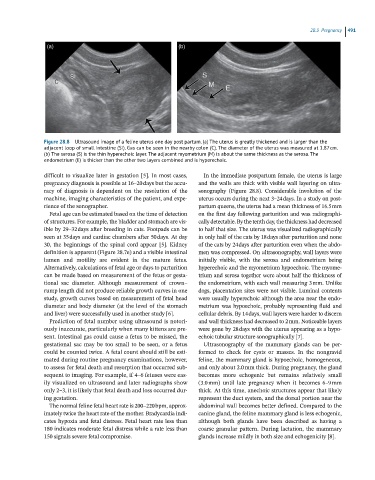
Figure 28.8 Ultrasound image of a feline uterus one day post partum. (a) The uterus is greatly thickened and is larger than the
adjacent loop of small intestine (SI). Gas can be seen in the nearby colon (C). The diameter of the uterus was measured at 1.87 cm.
(b) The serosa (S) is the thin hyperechoic layer. The adjacent myometrium (M) is about the same thickness as the serosa. The
endometrium (E) is thicker than the other two layers combined and is hyperechoic.
difficult to visualize later in gestation [5]. In most cases, In the immediate postpartum female, the uterus is large
pregnancy diagnosis is possible at 16–20 days but the accu and the walls are thick with visible wall layering on ultra
racy of diagnosis is dependent on the resolution of the sonography (Figure 28.8). Considerable involution of the
machine, imaging characteristics of the patient, and expe uterus occurs during the next 3–24 days. In a study on post
rience of the sonographer. partum queens, the uterus had a mean thickness of 16.5 mm
Fetal age can be estimated based on the time of detection on the first day following parturition and was radiographi
of structures. For example, the bladder and stomach are vis cally detectable. By the tenth day, the thickness had decreased
ible by 29–32 days after breeding in cats. Footpads can be to half that size. The uterus was visualized radiographically
seen at 35 days and cardiac chambers after 50 days. At day in only half of the cats by 18 days after parturition and none
30, the beginnings of the spinal cord appear [5]. Kidney of the cats by 24 days after parturition even when the abdo
definition is apparent (Figure 28.7e) and a visible intestinal men was compressed. On ultrasonography, wall layers were
lumen and motility are evident in the mature fetus. initially visible, with the serosa and endometrium being
Alternatively, calculations of fetal age or days to parturition hyperechoic and the myometrium hypoechoic. The myome
can be made based on measurement of the fetus or gesta trium and serosa together were about half the thickness of
tional sac diameter. Although measurement of crown– the endometrium, with each wall measuring 3 mm. Unlike
rump length did not produce reliable growth curves in one dogs, placentation sites were not visible. Luminal contents
study, growth curves based on measurement of fetal head were usually hyperechoic although the area near the endo
diameter and body diameter (at the level of the stomach metrium was hypoechoic, probably representing fluid and
and liver) were successfully used in another study [6]. cellular debris. By 14 days, wall layers were harder to discern
Prediction of fetal number using ultrasound is notori and wall thickness had decreased to 2 mm. Noticeable layers
ously inaccurate, particularly when many kittens are pre were gone by 28 days with the uterus appearing as a hypo
sent. Intestinal gas could cause a fetus to be missed, the echoic tubular structure sonographically [7].
gestational sac may be too small to be seen, or a fetus Ultrasonography of the mammary glands can be per
could be counted twice. A fetal count should still be esti formed to check for cysts or masses. In the nongravid
mated during routine pregnancy examinations, however, feline, the mammary gland is hypoechoic, homogeneous,
to assess for fetal death and resorption that occurred sub and only about 2.0 mm thick. During pregnancy, the gland
sequent to imaging. For example, if 4–6 fetuses were eas becomes more echogenic but remains relatively small
ily visualized on ultrasound and later radiographs show (3.0 mm) until late pregnancy when it becomes 6–9 mm
only 2–3, it is likely that fetal death and loss occurred dur thick. At this time, anechoic structures appear that likely
ing gestation. represent the duct system, and the dorsal portion near the
The normal feline fetal heart rate is 200–220 bpm, approx abdominal wall becomes better defined. Compared to the
imately twice the heart rate of the mother. Bradycardia indi canine gland, the feline mammary gland is less echogenic,
cates hypoxia and fetal distress. Fetal heart rate less than although both glands have been described as having a
180 indicates moderate fetal distress while a rate less than coarse granular pattern. During lactation, the mammary
150 signals severe fetal compromise. glands increase mildly in both size and echogenicity [8].