
Page 482 - Feline diagnostic imaging
P. 482
(a) (c)
(b) (d)
(e)
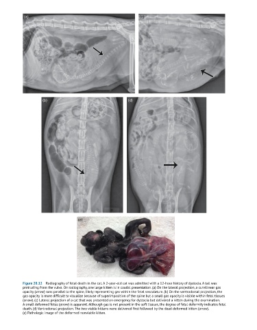
Figure 28.12 Radiography of fetal death in the cat. A 2-year-old cat was admitted with a 12-hour history of dystocia. A tail was
protruding from the vulva. On radiography, one large kitten is in caudal presentation. (a) On the lateral projection, a curvilinear gas
opacity (arrow) runs parallel to the spine, likely representing gas within the fetal vasculature. (b) On the ventrodorsal projection, the
gas opacity is more difficult to visualize because of superimposition of the spine but a small gas opacity is visible within fetal tissues
(arrow). (c) Lateral projection of a cat that was presented on emergency for dystocia but delivered a kitten during the examination.
A small deformed fetus (arrow) is apparent. Although gas is not present in the soft tissues, the degree of fetal deformity indicates fetal
death. (d) Ventrodorsal projection. The two viable kittens were delivered first followed by the dead deformed kitten (arrow).
(e) Pathologic image of the deformed nonviable kitten.