
Page 485 - Feline diagnostic imaging
P. 485
28.6 Diseisi of tsf seeas sep odu Dis Si se 497
(a)
(b) (c)
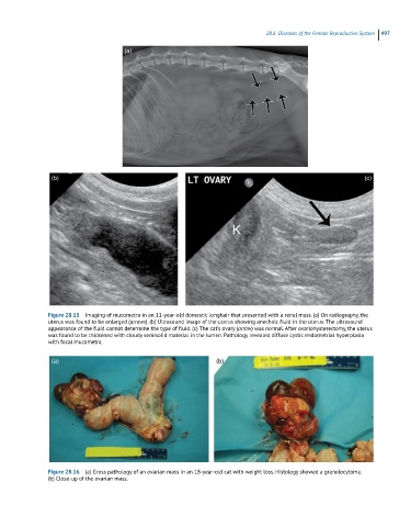
Figure 28.15 Imaging of mucometra in an 11-year-old domestic longhair that presented with a renal mass. (a) On radiography, the
uterus was found to be enlarged (arrows). (b) Ultrasound image of the uterus showing anechoic fluid in the uterus. The ultrasound
appearance of the fluid cannot determine the type of fluid. (c) The cat’s ovary (arrow) was normal. After ovariohysterectomy, the uterus
was found to be thickened with cloudy semisolid material in the lumen. Pathology revealed diffuse cystic endometrial hyperplasia
with focal mucometra.
(a) (b)
Figure 28.16 (a) Gross pathology of an ovarian mass in an 18-year-old cat with weight loss. Histology showed a granulocytoma.
(b) Close-up of the ovarian mass.