
Page 484 - Feline diagnostic imaging
P. 484
496 28 Reproduction
(a)
(b) (c)
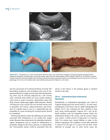
Figure 28.14 Pyometra in a 7-year-old domestic shorthair with a two-day history of vaginal discharge. Vaginal cytology showed
excessive neutrophils. (a) Transverse ultrasound image made from the ventral aspect of the abdomen. The horns of the uterus contain
cellular material (arrows). B, bladder. (b) Following ovariohysterectomy, purulent material was aspirated from the uterine horn. (c) Gross
pathology of the uterus of another cat, 8 years old, showing purulent material in the lumen.
pull the caudal pole of the ipsilateral kidney ventrally. The uterus in the female or the prostate gland or retained
descending duodenum and ascending colon may be dis testicle in the male.
placed medially by a right ovarian mass while the descend
ing colon may be similarly displaced by a left ovarian 28.6.4 Endometritis/Cystic Endometrial
mass. Anechoic areas within the mass usually represent
cystic areas or areas of necrosis. Hyperechoic areas with Hyperplasia
deep acoustic shadowing suggest mineralization. Masses Endometritis or endometrial hyperplasia can result in
involving the ovary include sex cord stromal tumors such irregular thickening of the uterine wall [11]. In some cases,
as granulosa cell tumors, adenocarcinomas, and terato enlargement of the uterus may be visible radiographically
mas. If a similar mass is seen in a cryptorchid tom, the (Figure 28.17), but evaluation for this condition is best
possibility of a mass involving the retained testicle should done using ultrasonography (Figures 28.17 and 28.18).
be considered. Small anechoic cysts are frequently present within the
Dorsocaudal masses within the abdomen are most often endometrial lining of the uterus, and the uterine lumen
associated with enlargement of the medial iliac lymph may contain a small amount of hypoechoic or anechoic
nodes. Because these nodes drain the hindlimbs and pelvic fluid. The ovaries may contain follicles or cysts. In the
region, enlargement may be caused by inflammatory or author’s experience, this condition is most often observed
neoplastic change in the uterus, prostate, or urinary blad in queens over the age of 5–6 years, but has been rarely
der. Ventrocaudal masses could be associated with the observed in younger cats. In more severe cases, the