
Page 486 - Feline diagnostic imaging
P. 486
498 28 Reproduction
hyperplastic endometrium may appear as a neoplastic Serial examination is useful to determine if involution is
mass within the uterine lumen (Figure 28.19). proceeding normally or not.
28.6.5 Failure of Involution 28.6.6 Cystic Ovaries
The uterus is normally enlarged following parturition. In A normal ovary is not appreciated on radiographs and is
the immediate postpartum period, the myometrium and better evaluated with ultrasound. A classic ovarian cyst is
endometrium will be visible as separate wall layers. anechoic and exhibits a distinct thin wall and deep acoustic
Compared to the nongravid appearance, the walls are enhancement. Small cysts may resemble follicles or cor
thicker, more irregular, and more echogenic. In one study pora lutea. Very large cysts are uncommon but may allow
of six cats, mean uterine diameter was 16 mm on day 1 fol the ovary to be seen on radiography, where it appears as a
lowing parturition. By 14 days, the uterine diameter had fluid opacity in or around the caudal pole of the kidney.
decreased to 6.2 mm, and by day 28, the uterus was still Cystic ovaries may occur in conjunction with pyometra,
identifiable on ultrasonography (mean uterine diameter cystic endometrial hyperplasia (Figures 28.17c and 28.18c),
4.7 mm) but wall layering was no longer observed [7]. or hydrometra. Cysts that produce hormones and affect
(a)
(b) (c)
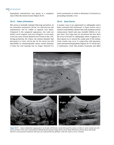
Figure 28.17 Cystic endometrial hyperplasia in an 8-year-old Persian cat. (a) Although the uterus is visible in some normal cats, the
uterus (arrows) in this cat is somewhat prominent. (b) Ultrasound image of the uterus shows thickening of the uterus and small
anechoic areas (arrows) consistent with cysts. (c) A cyst (arrow) was present in the left ovary. A, aorta; K, kidney.