
Page 488 - Feline diagnostic imaging
P. 488
500 28 Reproduction
(a) (b)
(c) (d)
(e) (f)
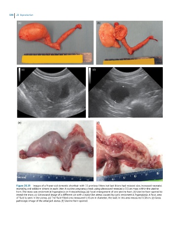
Figure 28.19 Images of a 9-year-old domestic shorthair with 11 previous litters but last litters had reduced size, increased neonatal
mortality, and stillborn kittens in each litter. A routine pregnancy check using ultrasound revealed a 3.5 cm mass within the uterine
horn. The mass was endometrial hyperplasia on histopathology. (a) Focal enlargement of one uterine horn. (b) Uterine horn opened to
reveal the mass. (c) Ultrasound image of a different cat with a mass-like uterus caused by cystic endometrial hyperplasia. A focal area
of fluid is seen in the uterus. (d) The fluid-filled area measured 1.43 cm in diameter; the wall in this area measured 0.38 cm. (e) Gross
pathologic image of the enlarged uterus. (f) Uterine horn opened.