
Page 493 - Feline diagnostic imaging
P. 493
29.3 ormal Spleen 505
capsule (Figure 29.3). They are similar in echogenicity to
surrounding fat and may not be visible on both sides. The
average length is 1.35 cm (range 0.5–2.33 cm) [2].
Sometimes a hyperechoic stripe is visible extending from
the hilus caudomedially caused by fat, fascia, lymphatics,
vessels, and glandular folds.
29.3 Normal Spleen
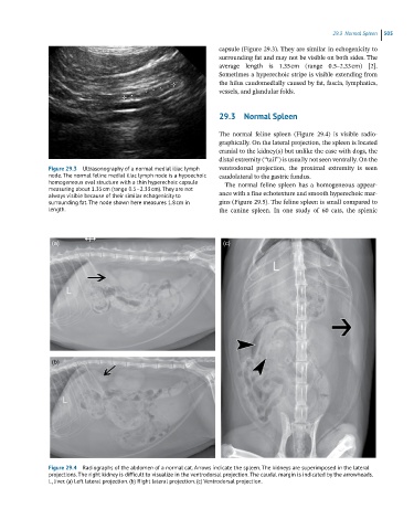
The normal feline spleen (Figure 29.4) is visible radio-
graphically. On the lateral projection, the spleen is located
cranial to the kidney(s) but unlike the case with dogs, the
distal extremity (“tail”) is usually not seen ventrally. On the
Figure 29.3 Ultrasonography of a normal medial iliac lymph ventrodorsal projection, the proximal extremity is seen
node. The normal feline medial iliac lymph node is a hypoechoic caudolateral to the gastric fundus.
homogeneous oval structure with a thin hyperechoic capsule The normal feline spleen has a homogeneous appear-
measuring about 1.35 cm (range 0.5–2.33 cm). They are not
always visible because of their similar echogenicity to ance with a fine echotexture and smooth hyperechoic mar-
surrounding fat. The node shown here measures 1.8 cm in gins (Figure 29.5). The feline spleen is small compared to
length. the canine spleen. In one study of 60 cats, the splenic
(a) (c)
(b)
Figure 29.4 Radiographs of the abdomen of a normal cat. Arrows indicate the spleen. The kidneys are superimposed in the lateral
projections. The right kidney is difficult to visualize in the ventrodorsal projection. The caudal margin is indicated by the arrowheads.
L, liver. (a) Left lateral projection. (b) Right lateral projection. (c) Ventrodorsal projection.