
Page 496 - Feline diagnostic imaging
P. 496
508 29 Hemolymphatic System
cranial mediastinum. If there is only pleural effusion and nodes to contain internal cysts while lymphomatous nodes
no mass, the lungs will become better inflated, decreasing were usually solid. Most thymomas were heterogeneous
the opacity in the cranial mediastinum. compared to 57% of lymphomatous nodes [8].
Ultrasonography can also be used to determine if a mass
is present and to further categorize any mass or masses that 29.4.3 Radiography of Abdominal Lymphoma
are visible. Ultrasound‐guided aspirates of the mass and
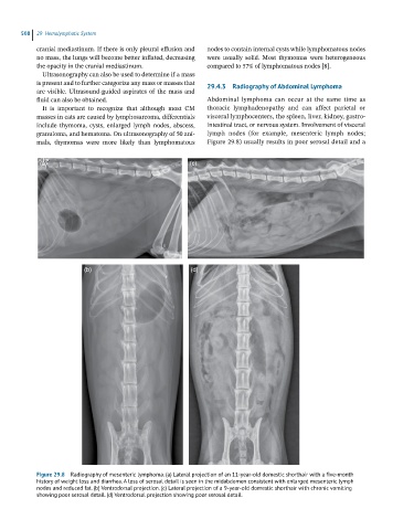
fluid can also be obtained. Abdominal lymphoma can occur at the same time as
It is important to recognize that although most CM thoracic lymphadenopathy and can affect parietal or
masses in cats are caused by lymphosarcoma, differentials visceral lymphocenters, the spleen, liver, kidney, gastro-
include thymoma, cysts, enlarged lymph nodes, abscess, intestinal tract, or nervous system. Involvement of visceral
granuloma, and hematoma. On ultrasonography of 50 ani- lymph nodes (for example, mesenteric lymph nodes;
mals, thymomas were more likely than lymphomatous Figure 29.8) usually results in poor serosal detail and a
(a) (c)
(b) (d)
Figure 29.8 Radiography of mesenteric lymphoma. (a) Lateral projection of an 11-year-old domestic shorthair with a five-month
history of weight loss and diarrhea. A loss of serosal detail is seen in the midabdomen consistent with enlarged mesenteric lymph
nodes and reduced fat. (b) Ventrodorsal projection. (c) Lateral projection of a 9-year-old domestic shorthair with chronic vomiting
showing poor serosal detail. (d) Ventrodorsal projection showing poor serosal detail.