
Page 497 - Feline diagnostic imaging
P. 497
29.4 The Many Mahees of nyymT yM 509
(b)
(a)
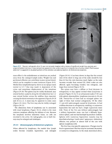
Figure 29.9 Thoracic radiographs of an 11-year-old domestic longhair with a history of significant weight loss, anorexia, and
suspected abdominal mass. A mass (arrows) was apparent in the caudal lung field. A complete loss of serosal detail (“fluid-opaque
abdomen”) is apparent in visualized portions of the abdomen. (a) Lateral projection. (b) Ventrodorsal projection.
mass effect in the midabdomen as intestines are pushed (Figure 29.14). It has been shown in dogs that the normal
away from the enlarged lymph nodes. Weight loss and ratio of the short to long axis of the node should be less
peritoneal effusion can contribute to poor serosal detail, than 0.5 but the ratio becomes much higher as the node
which can be complete in some instances (Figure 29.9). becomes rounder than normal [9]. Other nodes are also
Medial iliac lymphadenopathy shows as an opaque mass affected, again becoming rounder, less echogenic, and
ventral to L5–7 that may result in depression of the larger than normal (Figure 29.15).
colon and sometimes displacement of the intestines The spleen may have a diffuse or focal decrease in
(Figure 29.10). The spleen may be visibly thickened, may echogenicity or may appear normal when lymphoma is
extend further caudally along the left abdominal wall or present (Figure 29.16). In an ultrasound study of 101 cats
may extend further across the midline than expected, with splenic abnormalities, lymphoma was seen in 30 cats
causing the distal extremity to be apparent (Figures 29.10 [10]. Twenty‐five of these had an enlarged spleen but
and 29.11a–c). A mass may be apparent in some cases eight of those had normal echogenicity. Of the other
(Figure 29.11d,e). The liver may also be visibly enlarged 17 cats with splenomegaly caused by lymphoma, 16 had
(Figure 29.12). a diffuse decrease in echogenicity, mottling, or numerous
The alimentary form of lymphoma can be associated hypoechoic foci and one had a single hypoechoic mass
with renal lymphoma (Figure 29.13), which is the most [10]. Three of the five cats without splenomegaly had
common neoplasm affecting the kidneys of cats. Because a solitary hypoechoic mass, one had a diffusely hypo-
the neoplasm involves interstitial tissue, no cells are echoic spleen, and one had multiple hypoechoic nodules.
excreted in the urine. On radiography, one or both kid- Spleens with numerous hypoechoic nodules may be
neys may be enlarged and misshapen. described as having a “moth‐eaten” appearance. Abdominal
effusion was present in almost half of the cats with
splenic lymphoma [10].
29.4.4 Ultrasonography of Abdominal Lymphoma
The liver has a less specific appearance. Changes may be
When affected by lymphoma, the medial iliac lymph focal or generalized. The liver may be increased, decreased,
nodes become rounded, hypoechoic, and enlarged or normal in echogenicity. In the study mentioned above,