
Page 494 - Feline diagnostic imaging
P. 494
506 29 Hemolymphatic System
(a) (b)
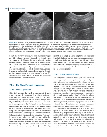
Figure 29.5 Ultrasonography of the normal feline spleen. The feline spleen is small compared to the canine spleen. Echogenicity is
homogeneous but unlike the canine spleen, the echogenicity of the feline spleen is more variable. The normal feline spleen is not
always hyperechoic but can be hypoechoic. (a) The spleen (S) is located in the near field with the stomach positioned cranially and
the colon positioned caudally. (b) Close-up of the feline spleen showing the hyperechoic capsule (arrowheads) and the location of the
splenic vein (arrow) as it exits the spleen. The larger splenic veins are visible within the spleen but the splenic arterial branches are
visible only with Doppler ultrasound. The stomach (St) is located cranially. The edge of the kidney is seen caudally.
height and width were measured from transverse images infiltration of lymph nodes by lymphoma can cause poor
as 8.2 ± 1.4 mm (mean, standard deviation) and lymphatic drainage that can cause a similar appearance.
26.7 ± 4.4 mm [3]. Whereas the canine spleen is consist- Radiographically, increased peribronchial and perivas-
ently hyperechoic, the feline spleen can be hypoechoic but cular opacity can cause blurring of pulmonary vessels.
still normal. Dogs have a sinusoidal spleen, allowing the Tracheobronchial lymphadenopathy usually appears as an
spleen to become large as a physiologic response. Cats have increase in perihilar opacity; the nodes are rarely visual-
a nonsinusoidal spleen [4], which does not readily enlarge ized as discrete opacities.
in response to physiologic changes. Extramedullary hemat-
opoiesis also seems to occur less frequently in cats [3].
Splenic veins are visible within the spleen but the smaller 29.4.2 Cranial Mediastinal Mass
arterial branches are not discernible. Cats that present with a CM mass (Figure 29.7) are usually
relatively young. In one study, the median age was 3 years.
Although in previous studies most affected cats were posi-
29.4 The Many Faces of Lymphoma tive for the feline leukemia virus (FeLV), 48/55 cats were
negative for FeLV and feline infectious peritonitis [7]. It was
thought that the change could be due to vaccination for
29.4.1 Thoracic Lymphoma
FeLV and removal of FeLV‐positive cats from cat colonies.
Often in lymphoma there will be enlargement of more A common presentation is dyspnea caused by compression
than one thoracic lymphocenter. On the lateral projection, of the trachea or coexisting pleural effusion. Lymphoma
there may be a discrete opacity on the floor of the thorax may invade the pericardium, heart, or cranial vena cava. The
representing enlargement of the sternal lymph node heart may also be indirectly affected because of compression
(Figure 29.6). Opacities near the trachea can occur second- of the lungs, vessels, or trachea. Lymphoma can be treated
ary to enlargement of the CM lymph nodes. The thymus with radiation or chemotherapy but recurrence is common
is located in the ventral portion of the cranial thorax and monitoring with ultrasonography can be helpful.
between the cranial lung lobes and may also be affected in On radiographs, the mass presents as a variably sized
lymphoma. Enlargement of the sternal or CM nodes or the opacity in the cranial mediastinum. Pericardial or pleural
thymus can cause a widening to the cranial mediastinum effusion may be present as well [5]. When there is a
to be apparent on the ventrodorsal projection. Pulmonary significant amount of pleural effusion, it can be difficult to
involvement may also occur with cellular infiltration determine whether or not a mass is present. Positional
appearing as an increase in pulmonary interstitial opacity radiography is useful in these cases. A radiograph is
although pulmonary infiltrates are more common in dogs exposed with the cat in an upright position. Gravity will
than in cats [5]. Lung lobe consolidation is rare but has cause the fluid to move to the lowest position. If a mass is
been reported secondary to lymphoma [6]. Additionally, present, there will continue to be fluid opacity in the