
Page 495 - Feline diagnostic imaging
P. 495
(a) (b)
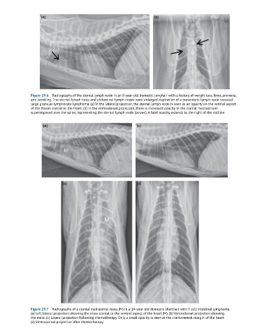
Figure 29.6 Radiography of the sternal lymph node in an 8-year-old domestic longhair with a history of weight loss, fever, anorexia,
and vomiting. The sternal lymph node and abdominal lymph nodes were enlarged. Aspiration of a mesenteric lymph node revealed
large granular lymphocyte lymphoma. (a) In the lateral projection, the sternal lymph node is seen as an opacity on the ventral aspect
of the thorax cranial to the heart. (b) In the ventrodorsal projection, there is increased opacity in the cranial mediastinum
superimposed over the spine, representing the sternal lymph node (arrows). A faint opacity extends to the right of the midline.
(a) (c)
(b) (d)
Figure 29.7 Radiography of a cranial mediastinal mass (M) in a 14-year-old domestic shorthair with T cell intestinal lymphoma.
(a) Left lateral projection showing the mass cranial to the ventral aspect of the heart (M). (b) Ventrodorsal projection showing
the mass. (c) Lateral projection following chemotherapy. Only a small opacity is seen at the cranioventral margin of the heart.
(d) Ventrodorsal projection after chemotherapy.