
Page 492 - Feline diagnostic imaging
P. 492
504 29 Hemolymphatic System
(b)
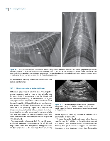
(a)
Figure 29.1 Radiographs of a 3-year-old domestic shorthair diagnosed with hemobartonellosis. The sternal lymph node (S) is mildly
enlarged, causing it to be visible radiographically. The approximate location of the tracheobronchial (TB) and cranial mediastinal (CM)
lymph nodes is indicated but these nodes are not enlarged. The sternal and cranial mediastinal lymph nodes are superimposed in the
ventrodorsal projection. (a) Lateral projection. (b) Ventrodorsal projection.
are located more caudally between the internal iliac and
median sacral arteries.
29.2.1 Ultrasonography of Abdominal Nodes
Abdominal lymphocenters are best seen with high‐fre-
quency transducers used in young or thin animals, with
the most visible lymphocenters being the jejunal and
medial iliac lymph nodes [2]. In the mesenteric group, jeju-
nal lymph nodes are long and oval with a reported mean of
20.1 mm (range 11.4–39.0 mm) [2]. They are usually some-
what hypoechoic although the center may be hyperechoic Figure 29.2 Ultrasonography of normal jejunal lymph node
(black arrow). The lymph nodes are long and oval. They are
compared to the periphery (Figure 29.2). They are not mainly hypoechoic but the center may be hyperechoic compared
always visualized, particularly in fat, older cats. Celiac lym- to the periphery.
phocenters have a similar appearance but are generally in
close proximity to an organ such as the spleen or liver. The various organs, watch for any evidence of abnormal celiac
caudal mesenteric and sacral lymph nodes are only found lymph nodes in the vicinity.
with difficulty [2]. To image the medial iliac lymph nodes, follow the aorta
When performing ultrasound, look for cranial mesen- caudally from the left kidney to the origin of the external
teric lymph nodes deep to the spleen on the left side and iliac arteries. Then, sweep the beam from ventrally and
deep to the duodenum on the right side. The jejunal nodes dorsally. The lymph nodes will appear as hypoechoic
will be near the root of the mesentery. When examining homogeneous oval structures with a thin hyperechoic