
Page 487 - Feline diagnostic imaging
P. 487
28.7 Diseisi of tsf eas sep odu Dis Si se 499
(a) (b)
(c)
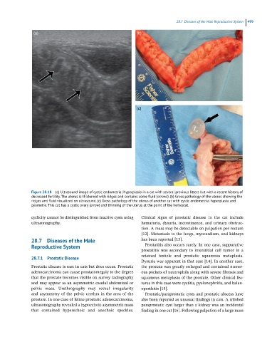
Figure 28.18 (a) Ultrasound image of cystic endometrial hyperplasia in a cat with several previous litters but with a recent history of
decreased fertility. The uterus is thickened with ridges and contains some fluid (arrows). (b) Gross pathology of the uterus showing the
ridges and fluid visualized on ultrasound. (c) Gross pathology of the uterus of another cat with cystic endometrial hyperplasia and
pyometra. This cat has a cystic ovary (arrow) and thinning of the uterus at the point of the hemostat.
cyclicity cannot be distinguished from inactive cysts using Clinical signs of prostatic disease in the cat include
ultrasonography. hematuria, dysuria, incontinence, and urinary obstruc
tion. A mass may be detectable on palpation per rectum
[12]. Metastasis to the lungs, myocardium, and kidneys
28.7 Diseases of the Male has been reported [13].
Reproductive System Prostatitis also occurs rarely. In one case, suppurative
prostatitis was secondary to interstitial cell tumor in a
28.7.1 Prostatic Disease retained testicle and prostatic squamous metaplasia.
Dysuria was apparent in that case [14]. In another case,
Prostatic disease is rare in cats but does occur. Prostatic the prostate was greatly enlarged and contained numer
adenocarcinoma can cause prostatomegaly to the degree ous pockets of neutrophils along with severe fibrosis and
that the prostate becomes visible on survey radiography squamous metaplasia of the prostate. Other clinical fea
and may appear as an asymmetric caudal abdominal or tures in this case were cystitis, pyelonephritis, and balan
pelvic mass. Urethrography may reveal irregularity oposthitis [15].
and asymmetry of the pelvic urethra in the area of the Prostatic/paraprostatic cysts and prostatic abscess have
prostate. In one case of feline prostatic adenocarcinoma, also been reported as unusual findings in cats. A trilobed
ultrasonography revealed a hypoechoic asymmetric mass paraprostatic cyst larger than a kidney was an incidental
that contained hyperechoic and anechoic speckles. finding in one cat [16]. Following palpation of a large mass