
Page 483 - Feline diagnostic imaging
P. 483
28.6 Diseisi of tsf seeas sep odu Dis Si se 495
(a) (b) (c)
(d) (e)
(f) (g) (h)
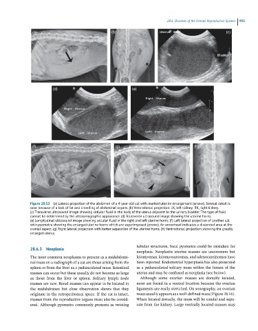
Figure 28.13 (a) Lateral projection of the abdomen of a 4-year-old cat with marked uterine enlargement (arrows). Serosal detail is
poor because of a lack of fat and crowding of abdominal organs. (b) Ventrodorsal projection. LK, left kidney; RK, right kidney.
(c) Transverse ultrasound image showing cellular fluid in the body of the uterus adjacent to the urinary bladder. The type of fluid
cannot be determined by the ultrasonographic appearance. (d) Transverse ultrasound image showing the uterine horns.
(e) Longitudinal ultrasound image showing cellular fluid in the right and left uterine horns. (f) Left lateral projection of another cat
with pyometra showing the enlarged uterine horns which are superimposed (arrows). An arrowhead indicates a distended area at the
cranial aspect. (g) Right lateral projection with better separation of the uterine horns. (h) Ventrodorsal projection showing the greatly
enlarged uterus.
tubular structures, focal pyometra could be mistaken for
28.6.3 Neoplasia neoplasia. Neoplastic uterine masses are uncommon but
The most common neoplasms to present as a midabdomi leiomyomas, leiomyosarcomas, and adenocarcinomas have
nal mass on a radiograph of a cat are those arising from the been reported. Endometrial hyperplasia has also presented
spleen or from the liver as a pedunculated mass. Intestinal as a pedunculated solitary mass within the lumen of the
masses can occur but these usually do not become as large uterus and may be confused as neoplasia (see below).
as those from the liver or spleen. Solitary lymph node Although some ovarian masses are dorsally located,
masses are rare. Renal masses can appear to be located in most are found in a ventral location because the ovarian
the midabdomen but close observation shows that they ligaments are easily stretched. On sonography, an ovarian
originate in the retroperitoneal space. If the cat is intact, mass usually appears as a well‐defined mass (Figure 28.16).
masses from the reproductive organs must also be consid When located dorsally, the mass will be caudal and sepa
ered. Although pyometra commonly presents as twisting rate from the kidney. Large ventrally located masses may