
Page 498 - Feline diagnostic imaging
P. 498
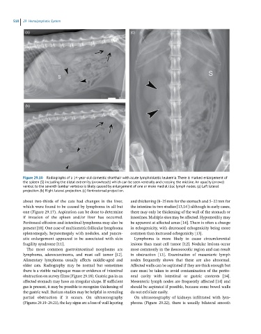
510  29  Hemolymphatic System

             (a)                                              (c)


             (b)


            Figure 29.10  Radiographs of a 14-year-old domestic shorthair with acute lymphoblastic leukemia. There is marked enlargement of
            the spleen (S) including the distal extremity (arrowheads) which can be seen ventrally and crossing the midline. An opacity (arrows)
            ventral to the seventh lumbar vertebra is likely caused by enlargement of one or more medial iliac lymph nodes. (a) Left lateral
            projection. (b) Right lateral projection. (c) Ventrodorsal projection.

            about  two‐thirds  of  the  cats  had  changes  in  the  liver,   and thickening (8–25 mm for the stomach and 5–22 mm for
            which were found to be caused by lymphoma in all but   the intestine in two studies [13,14]) although in early cases,
            one (Figure 29.17). Aspiration can be done to determine   there may only be thickening of the wall of the  stomach or
            if  invasion  of  the  spleen  and/or  liver  has  occurred.   intestines. Multiple sites may be affected. Hypomotility may
            Peritoneal effusion and intestinal lymphoma may also be   be apparent at affected areas [14]. There is often a change
            present [10]. One case of multicentric follicular lymphoma   in echogenicity, with decreased echogenicity being more
            splenomegaly, hepatomegaly with nodules, and pancre-  common than increased echogenicity [13].
            atic  enlargement  appeared  to  be  associated  with  skin   Lymphoma  is  more  likely  to  cause  circumferential
            fragility syndrome [11].                          lesions than mast cell tumor [12]. Nodular lesions occur
              The  most  common  gastrointestinal  neoplasms  are   most commonly in the ileocecocolic region and can result
            lymphoma,  adenocarcinoma,  and  mast  cell  tumor  [12].   in  obstruction  [13].  Examination  of  mesenteric  lymph
            Alimentary  lymphoma  usually  affects  middle‐aged  and   nodes  frequently  shows  that  these  are  also  abnormal.
            older  cats.  Radiography  may  be  normal  but  sometimes   Affected walls can be aspirated if they are thick enough but
            there is a visible radiopaque mass or evidence of intestinal   care must be taken to avoid contamination of the perito-
            obstruction on survey films (Figure 29.18). Gastric gas in an   neal  cavity  with  intestinal  or  gastric  contents  [14].
            affected stomach may have an irregular shape. If sufficient   Mesenteric lymph nodes are frequently affected [14] and
            gas is present, it may be possible to recognize thickening of   should be aspirated if possible, because some bowel walls
            the gastric wall. Barium studies may be helpful in revealing   do not exfoliate easily.
            partial  obstruction  if  it  occurs.  On  ultrasonography   On  ultrasonography  of  kidneys  infiltrated  with  lym-
            (Figures 29.19–29.21), the key signs are a loss of wall layering   phoma  (Figure  29.22),  there  is  usually  bilateral  smooth