
Page 499 - Feline diagnostic imaging
P. 499
29.4 The Many Mahees of nyymT yM 511
(a) (c)
(b)
(e)
(d)
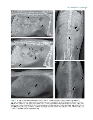
Figure 29.11 Radiography of enlarged spleen (S) in an 11-year-old domestic shorthair diagnosed with lymphoma following
aspiration of the bone marrow. Margins of the spleen are indicated by the arrowheads. The proximal extremity of the spleen is more
difficult to visualize in the right lateral projection because of superimposition of the kidneys indicated by small arrows. (a) Left lateral
projection. (b) Right lateral projection. (c) Ventrodorsal projection. (d) Lateral projection of referral radiography of a cat with a splenic
mass determined to be a high-grade B cell lymphoma following splenectomy. There is an opacity (arrowheads) in the area of the distal
extremity of the spleen. (e) Ventrodorsal projection.