
Page 504 - Feline diagnostic imaging
P. 504
516 29 Hemolymphatic System
(b)
(a)
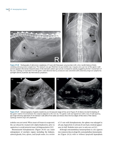
Figure 29.18 Radiography of alimentary lymphoma. A 9-year-old Himalayan cat presented with a two-month history of daily
vomiting and progressive weight loss. The stomach was gas filled but the gas bubble was irregular and a portion of the gastric wall
(arrows) appears thickened. Adjacent areas of the stomach contain gas but are increased in opacity, suggesting further involvement of
the wall. Cytology on aspirates from the gastric wall obtained during ultrasound was consistent with alimentary large cell lymphoma.
(a) Right lateral projection. (b) Ventrodorsal projection.
(a) (b)
Figure 29.19 Ultrasonography of gastric lymphoma. (a) Ultrasound image of the cat in Figure 29.18 shows marked thickening of
the gastric wall (1.36 cm between the calipers) adjacent to the liver (L). The gastric wall was hypoechoic with a loss of wall layering.
(b) Image showing aspiration of the stomach wall (SW) of the same cat. Arrows show the two edges of the bevel of the needle.
Cytology showed large cell lymphoma.
nodules was not noted. When mast cell tumor is suspected, of 15 cats with histoplasmosis, the spleen was enlarged in
the cat should be treated with diphenhydramine prior to all cats, hypoechoic in almost all and had a mottled appear-
aspiration to avoid potential mast cell degranulation [21]. ance in half. Nodules were seen in only one cat [22].
Disseminated histoplasmosis (Figure 29.24) can cause Although extramedullary hematopoiesis in cats appears
enlargement of multiple organs including the kidneys, less common than in dogs [4], extramedullary hematopoie-
adrenal glands, liver, spleen, and lymph nodes. In a review sis (Figure 29.25) with or without lymphoid hyperplasia