
Page 507 - Feline diagnostic imaging
P. 507
29.5 DesheMeshees of Thef mphehea 519
(a) (b)
(c)
(d)
(e)
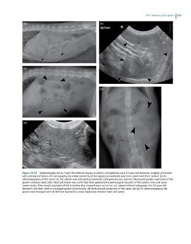
Figure 29.23 Splenomegaly due to mast cell without change in splenic echogenicity. (a) A 17-year-old domestic longhair presented
with a history of ataxia. On radiography, the distal extremity of the spleen (arrowheads) was more prominent than normal. (b) On
ultrasonography of the same cat, the spleen was enlarged (arrowheads). Echogenicity was normal. Ultrasound-guided aspiration of the
spleen revealed mast cells. Mast cell tumor was confirmed after splenectomy and surgical biopsies of the spleen, liver, and cecal
lymph nodes. Fine needle aspirates of the intestine also showed mast cell tumor. (c) Lateral referral radiograph of a 16-year-old
domestic shorthair with an enlarged spleen (arrowheads). (d) Ventrodorsal projection of the same cat. (e) On ultrasonography, the
spleen was enlarged with ill-defined hypoechoic areas. Aspiration showed mast cell tumor.