
Page 509 - Feline diagnostic imaging
P. 509
29.5 DesheMeshees of Thef mphehea 521
(a)
(c) (b)
(d)
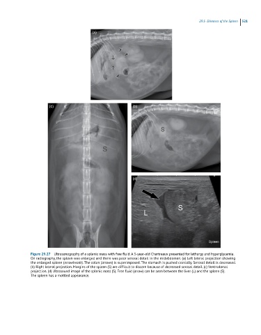
Figure 29.27 Ultrasonography of a splenic mass with free fluid. A 5-year-old Chartreaux presented for lethargy and hyperglycemia.
On radiography, the spleen was enlarged and there was poor serosal detail in the midabdomen. (a) Left lateral projection showing
the enlarged spleen (arrowheads). The colon (arrows) is superimposed. The stomach is pushed cranially. Serosal detail is decreased.
(b) Right lateral projection. Margins of the spleen (S) are difficult to discern because of decreased serosal detail. (c) Ventrodorsal
projection. (d) Ultrasound image of the splenic mass (S). Free fluid (arrow) can be seen between the liver (L) and the spleen (S).
The spleen has a mottled appearance.