
Page 505 - Feline diagnostic imaging
P. 505
29.5 DesheMeshees of Thef mphehea 517
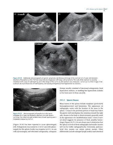
(a) (b)
(c)
Figure 29.20 Additional ultrasonography of gastric lymphoma. (a) Ultrasound image of the stomach of a 9-year-old domestic
shorthair, FIV positive on the FIV/FeLV snap test and diagnosed with large cell lymphoma. The wall of the stomach is severely
thickened with a loss of wall layering. (b) In this image of the same cat, the stomach wall measures 1.67 cm. (c) In a third image of the
stomach, the wall shows loss of wall layering and varying echogenicity from hyperechoic to anechoic.
change usually consisted of decreased echogenicity, focal
hypoechoic nodules, or mottling but hyperechoic nodules
or foci were seen in three cats [10].
29.5.2 Splenic Masses
Mass lesions of the spleen include neoplasia (particularly
hemangiosarcoma) and hematoma. The appearance on
radiography varies with the location of the mass in the
spleen. A mass in the proximal portion or head may indent
Figure 29.21 Ultrasonography of lymphoma in the small the gastric wall and displace the intestines toward the right
intestine of a 9-year-old domestic shorthair cat with chronic side. Masses in the body or distal extremity generally result
vomiting. The intestinal wall exhibits loss of wall layering and is in the appearance of a midabdominal mass. A loss of sero-
thickened, measuring 0.67 cm. sal detail suggests the presence of fluid, possibly hemor-
rhage (Figure 29.27). It is not always easy to determine that
(Figure 29.26) has been reported to cause splenomegaly the spleen is involved from radiographs and smaller masses
[10]. Echogenicity was normal in 11 of 27 cats with spleno- will not be visible radiographically. Conversely, peduncu-
megaly but the splenic border was irregular in 8/11. In cats lated liver masses can mimic splenic masses. Other
with splenomegaly and abnormal echogenicity, echogenic differentials include enlarged lymph node(s) and intestinal