
Page 510 - Feline diagnostic imaging
P. 510
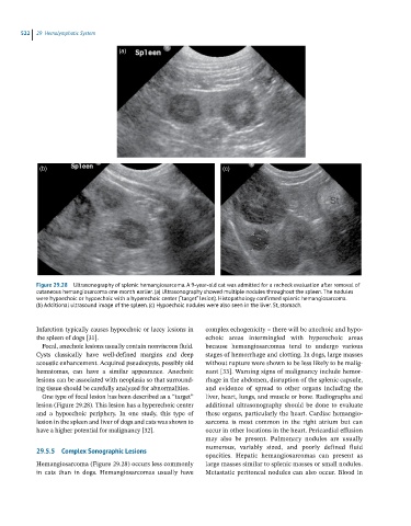
522  29  Hemolymphatic System

                                    (a)


             (b)                                                   (c)


            Figure 29.28  Ultrasonography of splenic hemangiosarcoma. A 9-year-old cat was admitted for a recheck evaluation after removal of
            cutaneous hemangiosarcoma one month earlier. (a) Ultrasonography showed multiple nodules throughout the spleen. The nodules
            were hypoechoic or hypoechoic with a hyperechoic center (“target” lesion). Histopathology confirmed splenic hemangiosarcoma.
            (b) Additional ultrasound image of the spleen. (c) Hypoechoic nodules were also seen in the liver. St, stomach.


            Infarction typically causes hypoechoic or lacey lesions in     complex echogenicity – there will be anechoic and hypo-
            the spleen of dogs [31].                          echoic  areas  intermingled  with  hyperechoic  areas
              Focal, anechoic lesions usually contain nonviscous fluid.   because  hemangiosarcomas  tend  to  undergo  various
            Cysts  classically  have  well‐defined  margins  and  deep   stages of hemorrhage and clotting. In dogs, large masses
            acoustic enhancement. Acquired pseudocysts, possibly old   without rupture were shown to be less likely to be malig -
            hematomas,  can  have  a  similar  appearance.  Anechoic   nant [33]. Warning signs of malignancy include hemor-
            lesions can be associated with neoplasia so that surround-  rhage in the abdomen, disruption of the splenic capsule,
            ing tissue should be carefully analyzed for abnormalities.  and  evidence  of  spread  to  other  organs  including  the
              One type of focal lesion has been described as a “target”   liver, heart, lungs, and muscle or bone. Radiographs and
            lesion (Figure 29.28). This lesion has a hyperechoic center   additional  ultrasonography  should  be  done  to  evaluate
            and  a  hypoechoic  periphery.  In  one  study,  this  type  of   these organs, particularly the heart. Cardiac hemangio-
            lesion in the spleen and liver of dogs and cats was shown to   sarcoma  is  most  common  in  the  right  atrium  but  can
            have a higher potential for malignancy [32].      occur in other locations in the heart. Pericardial effusion
                                                              may  also  be  present.  Pulmonary  nodules  are  usually
                                                              numerous,  variably  sized,  and  poorly  defined  fluid
            29.5.5  Complex Sonographic Lesions
                                                                opacities.  Hepatic  hemangiosarcomas  can  present  as
            Hemangiosarcoma (Figure 29.28) occurs less commonly   large masses similar to splenic masses or small nodules.
            in  cats  than  in  dogs.  Hemangiosarcomas  usually  have   Metastatic peritoneal nodules can also occur. Blood in