
Page 512 - Feline diagnostic imaging
P. 512
524 29 Hemolymphatic System
29.6 Other Diseases of Lymph Lymphoid hyperplasia (Figure 29.32) can occur secondary
Nodes and Lymphatics to infection in other organs by disease such as cryptococ-
cosis [34]. These nodes can be enlarged, round, and hypo-
Enlargement and changes in shape or echogenicity of echoic similar to the appearance seen with lymphoma but
lymph nodes are most frequently caused by lymphoma in lymphoma should always be considered when nodes have
cats but can have other causes. Reactive lymphadenopathy this appearance.
can occur because of disease in the gastrointestinal tract or Lymph nodes that are enlarged secondary to metastatic
other organs (Figure 29.31). Usually in these cases, the disease may have increased or decreased echogenicity
lymph nodes are larger or more prominent than usual and often have irregular margins or are misshapen.
but echogenicity, size, and shape are close to normal. Local lymph nodes should always be examined when a
neoplasm is confirmed or suspected (Figure 29.33).
Heterogeneous lymph nodes in dogs and humans are
more likely to be malignant than benign but in one study,
this association was not seen in cats. Nonetheless, the
discovery of misshapen lymph nodes with altered echo-
genicity should prompt a search for neoplasia in the area
drained by the abnormal node(s) [35]. Lymph nodes can
be aspirated to determine if there is primary disease such
as lymphoma or metastatic disease. Although lymphoma
usually results in round, enlarged, hypoechoic nodes,
occasionally lymph nodes can be distorted with abnor-
mal echogenicity when affected by lymphoma.
Lymph nodes can also become primarily infected.
Infected nodes may be just enlarged (Figure 29.34) or may
become abscessed. Abscessed lymph nodes (Figure 29.34)
may obviously contain fluid or may appear to contain
Figure 29.31 Ultrasonography of reactive lymphadenopathy. A hypoechoic nodules that yield purulent material on
12-year-old domestic shorthair had a history of chronic vomiting aspiration.
and weight loss. On ultrasonography, small intestinal wall Neoplasia can also affect the lymphatic endothelium.
layering was normal but the wall was mildly thickened,
suggesting inflammatory bowel disease. The mesenteric lymph Lymphangiosarcoma was reported in two young cats that
nodes were mildly enlarged consistent with reactive presented with petechiae, ecchymoses, lymphoedema, and
lymphadenopathy. serosanguinous discharge [36]. One cat also had hemolytic
(a) (b)
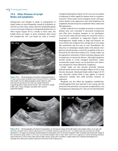
Figure 29.32 Ultrasonography of lymphoid hyperplasia. (a) Ultrasound image of the left medial iliac lymph node in an 8-year-old
domestic shorthair with a colonic adenocarcinoma. Subtotal colectomy was performed. The lymph node was rounded and mildly
decreased in echogenicity. Cytology showed lymphoid hyperplasia with no obvious neoplastic cells. B, bladder. (b) Ultrasound image of
the mesenteric lymph nodes in a 4.5-year-old domestic longhair with a colon mass found to be suppurative colitis following resection
and anastomosis of the colon. Eight months after colonic surgery, the mesenteric lymph nodes were enlarged and hypoechoic.
Cytology after ultrasound-guided aspiration showed lymphoid hyperplasia.