
Page 477 - Feline diagnostic imaging
P. 477
(a) (b)
(d)
(c)
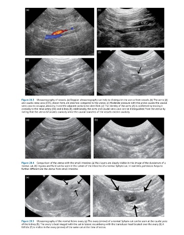
Figure 28.3 Ultrasonography of vessels. (a) Doppler ultrasonography can help to distinguish the uterus from vessels. (b) The aorta (A)
and caudal vena cava (CVC), shown here, are anechoic compared to the uterus. (c) Moderate pressure with the probe causes the caudal
vena cava to collapse, allowing it and the adjacent aorta to be identified. (d) The identity of the aorta (A) is confirmed by tracing it
cranially to the renal artery (RA) and kidney (K). Additionally, the aorta and caudal vena cava can be distinguished from the uterus by
noting that the uterus bifurcates cranially while the caudal branches of the vessels extend caudally.
(a) (b)
Figure 28.4 Comparison of the uterus with the small intestine. (a) Wall layers are clearly visible in this image of the duodenum of a
normal cat. (b) Ingesta and fluid can be seen in the lumen of the intestine of a normal Sphynx cat. In real time, peristalsis helps to
further differentiate the uterus from small intestine.
(a) (b)
Figure 28.5 Ultrasonography of the normal feline ovary. (a) The ovary (arrows) of a normal Sphynx cat can be seen at the caudal pole
of the kidney (K). The ovary is best imaged with the cat in lateral recumbency with the transducer head located over the ovary. (b) A
follicle (F) is visible in the ovary (arrows) of the same cat at the time of estrus.