
Page 471 - Feline diagnostic imaging
P. 471
27.5 Urethra 483
(a) (b)
Figure 27.34 Ultrasonography of lymphosarcoma in the urinary bladder of a 6-year-old castrated male cat with progressive weight
loss. There was a medullary rim sign in each of the kidneys (Figure 27.4b). (a) There was marked thickening of the bladder wall at the
apex with a loss of wall layering. (b) The bladder wall measured between 0.7 and 0.9 cm.
results less often in cats than in dogs because feline cystic the pelvic bones or gunshot. On positive contrast urethrog-
neoplasia is more common at the apex than at the trigone. raphy, contrast is seen in the periurethral tissues, confirm-
On ultrasonography, the location of the mass (trigone or ing that rupture has occurred.
apex), extent of bladder involvement, number of involved
wall layers, and presence of metastasis should be evaluated.
Very little lumen is visible in some cases. The urethra should 27.5.2 Urethral Calculi
be examined to see if there is any extension distally. Some urethral calculi are radiopaque and can be seen on
survey radiography (Figure 27.35). Urethrography can be
27.5 Urethra used to identify radiolucent calculi, which will appear as a
filling defect in the contrast column. Calculi are usually
asymmetrically located with irregular shapes and irregu-
27.5.1 Ruptured Urethra
lar, indistinct margins. Larger calculi may distend the ure-
Rupture of the urethra can cause increased opacity and thra. Filling defects can also be caused by blood clots,
loss of detail of structures in the pelvic region [44]. which may have a similar appearance but which will not
There may also be evidence of trauma such as fracture of distend the urethra. Air bubbles will also not distend the
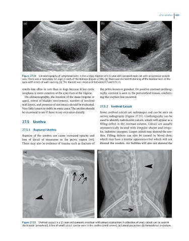
(a) (b)
Figure 27.35 Urethral calculi in a 15-year-old domestic shorthair with urinary obstruction. A collection of small calculi can be seen in
the bladder (arrowhead). A line of small calculi can be seen in the urethra (small arrows). (a) Lateral projection. (b) Ventrodorsal projection.