
Page 469 - Feline diagnostic imaging
P. 469
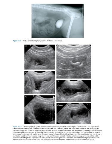
Figure 27.31  Double contrast cystography showing thickened bladder wall.

                            (a)                                (b)


                            (c)


                                                               (d)


                            (e)


                                                               (f)


               Figure 27.32  Ultrasonography of cystitis. (a) Ultrasound image of a 10-year-old domestic longhair presented with a one-day history of
               hematuria. The bladder wall is thickened at 0.84 cm. (b) Hyperechoic material is seen in the lumen of the bladder of the cat in (a). (c) An
               ultrasound image of a 12-year-old castrated male cat shows focal thickening of the bladder wall measuring 1.01 cm long and 0.94 cm deep.
               Ultrasound-guided aspiration can be done when there is concern for neoplasia. (d) In other cases, thickening is more uniform, as shown in
               this image of another cat with cystitis. (e) An ultrasound scan of a 9-year-old cat with cystitis shows a thickened bladder with a small cyst in
               the bladder wall. (f) In this ultrasound image of a 14-year-old Himalayan cat made with the patient standing, echogenic cellular debris
               could be seen drifting to the dependent side of the urinary bladder. Echogenic cellular material is a common finding even in normal cats
               and may be attributed to fat in some cases. The urinary bladder was not thickened and there was no clinical evidence of cystitis.