
Page 467 - Feline diagnostic imaging
P. 467
(a)
(b) (c)
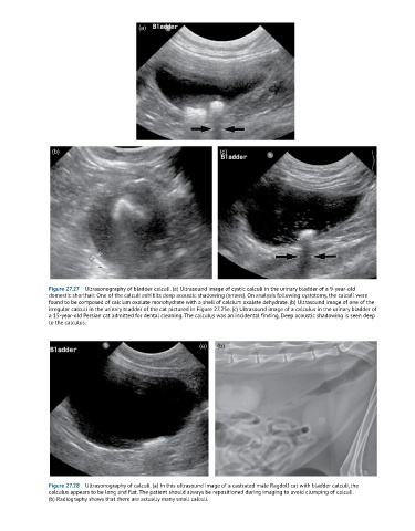
Figure 27.27 Ultrasonography of bladder calculi. (a) Ultrasound image of cystic calculi in the urinary bladder of a 9-year-old
domestic shorthair. One of the calculi exhibits deep acoustic shadowing (arrows). On analysis following cystotomy, the calculi were
found to be composed of calcium oxalate monohydrate with a shell of calcium oxalate dehydrate. (b) Ultrasound image of one of the
irregular calculi in the urinary bladder of the cat pictured in Figure 27.25e. (c) Ultrasound image of a calculus in the urinary bladder of
a 15-year-old Persian cat admitted for dental cleaning. The calculus was an incidental finding. Deep acoustic shadowing is seen deep
to the calculus.
(a) (b)
Figure 27.28 Ultrasonography of calculi. (a) In this ultrasound image of a castrated male Ragdoll cat with bladder calculi, the
calculus appears to be long and flat. The patient should always be repositioned during imaging to avoid clumping of calculi.
(b) Radiography shows that there are actually many small calculi.