
Page 446 - Small Animal Clinical Nutrition 5th Edition
P. 446
460 Small Animal Clinical Nutrition
istration (by pump or gravity flow) of food into the stomach.
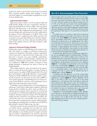
Box 25-5. Nasoesophageal Tube Placement.
VetBooks.ir Most veterinary patients tolerate bolus feedings of enteral Nasoesophageal tubes are generally used for three to seven days,
nutritional support via nasoesophageal, esophagostomy or gas-
trostomy feeding tubes.
but are occasionally used longer (weeks if moved to the opposite
JEJUNOSTOMY TUBES side every seven days). Polyurethane tubes (6 to 8 Fr., 90 to 100
cm) with or without a weighted tip and silicone feeding tubes (3.5
Jejunostomy tubes (J-tubes, 5 to 8 Fr.) are placed within the
to 10 Fr., 20 to 105 cm) may be placed in the caudal esophagus
small intestine, ideally at the time of exploratory celiotomy, to
or stomach. The preferred placement of all tubes originating cra-
bypass the proximal GI tract (Orton, 1986). J-tubes may also be
nial to the stomach is in the caudal esophagus to minimize gastric
placed by mini-laparotomy, or by threading a small feeding
reflux and subsequent esophagitis. An 8-Fr. tube will pass through
tube through a larger esophagostomy, pharyngostomy or gas- the nasal cavity of most dogs; a 5- Fr. tube is more comfortable for
trostomy feeding tube and placing the tip of the smaller tube in cats.
the jejunum (Crowe, 1986; Jergens et al, 2007). There is risk, The length of tube to be inserted is determined by measuring
however, that even a weighted-tip tube will be returned to the from the nasal planum along the side of the animal to the caudal
stomach by reverse peristalsis. Ideally, food should be adminis- margin of the last rib (Figure 1) and marking the tube at a point
tered through J-tubes at a slow, continuous drip delivered by a that is approximately three-fourths of the total measured length
pump. Some patients, however, will tolerate frequent small- with a piece of adhesive tape or an indelible marker. This mark is
how far the tube should be inserted. Tape will also provide a tab to
bolus feedings.
secure the tube. The animal’s nose is desensitized by placing a
few drops of topical anesthetic (2% lidocaine or 0.5% propara-
Amount to Feed and Feeding Schedule
caine) into a nostril and tilting the head upward for a few seconds.
Feeding plans require an understanding of the patient’s meta-
The tip of the tube is lubricated with a water-soluble lubricant or 2
bolic state relative to changes in metabolism resulting from to 5% lidocaine ointment/jelly before passage.
ongoing food deprivation. Estimating a patient’s approximate To pass the tube, direct the tip in a caudoventral, medial direc-
caloric requirement is important because feeding more of any tion into the ventrolateral aspect of the external nares. The head is
food than is necessary may cause metabolic complications. generally held in a normal static position. As soon as the tip of the
Overfeeding patients is possible through a feeding tube and catheter reaches the medial septum at the floor of the nasal cavi-
should be avoided because it results in metabolic and mechan- ty in dogs, the external nares are pushed dorsally, which opens the
ical complications. Table 25-7 provides an example of using ventral meatus, ensuring passage of the tube into the oropharynx
(Figure 2). To aid passage, the proximal end of the tube is lifted
feeding guidelines to determine how much to feed and the
as the nose is pushed upward (Figure 2). In cats, because of the
feeding schedule.
lack of a well-developed alar fold, the tube can be inserted initial-
The feeding schedule is often determined by the patient’s
ly in a ventromedial direction and continued directly into the
ability to tolerate food and the logistics of feeding. Feeding an
oropharynx. The tube is inserted until the adhesive tape tab or
amount equal to the patient’s RER during the first 24 hours of indelible mark is reached (Figure 3).
food reintroduction, if physically tolerated, is recommended. To evaluate proper tube placement, 3 to 15 ml of sterile water
Feeding one-third of RER the first 24 hours and then increas- or saline solution may be injected through the tube and the animal
ing the amount by one-third every 24 hours until at RER is a evaluated for coughing (Figure 4). A lateral radiograph may be
more cautious approach to initial feeding, but is not always nec- taken of the neck to confirm the tube is placed in the caudal
essary. Foods should be warmed to room temperature, but not esophagus (i.e., over the larynx). After confirmation of position, the
higher than body temperature, before feeding. tube is secured with either sutures or glue. The first tape tab is
secured to the skin just lateral to the external nares.A second tape
Food boluses must be infused slowly (over approximately one
tab is secured to the skin on the dorsal nasal midline, just rostral
minute per 5 ml of food) to allow gastric expansion. Daily food
to the level of the eyes. An Elizabethan collar is used in most ani-
dosage should be divided into several meals according to the
mals to prevent inadvertent removal of the tube (Figure 5).
expected stomach capacity. Gastric capacities for cats and dogs
Complications of nasoesophageal intubation include epistaxis,
are typically 5 to 10 ml/kg body weight during initial food rein- lack of tolerance of the procedure and inadvertent removal of the
troduction.Maximum capacities as high as 45 to 90 ml/kg body tube by the animal. Incidence of tube removal by the animal has
weight have been measured in cats and dogs when fully re-ali- been reported to be as high as 50% even with use of collars.
mented. Most often, the patient’s RER can be met in volumes Nasoesophageal tubes should not be used in vomiting patients or
far less than these maximum gastric capacities. Salivating, gulp- those with respiratory disease.
ing, retching and vomiting may occur when too much food has
been infused or when the infusion rate is too fast.
Research in people has demonstrated that the stomach does
not “shrink” during a prolonged fast, but rather the stretch
receptors are more sensitive and stimulated by a smaller volume
when refeeding occurs. Feeding should be stopped at the first
sign of retching or salivating; then the meal size reduced by
50% for 24 hours and then increased by 25% gradually. Foods