
Page 449 - Small Animal Clinical Nutrition 5th Edition
P. 449
Enteral-Assisted Feeding 463
VetBooks.ir Several techniques have been described for mid-cervical placement
Box 25-7. Esophagostomy Tube Placement.
of esophagostomy tubes in dogs and cats. The animal receives light
general anesthesia for esophagostomy tube placement.The entire lat-
eral cervical region from the ventral midline to near the dorsal midline
is clipped and aseptically prepared for surgery.
In one technique, appropriately sized, curved Kelly, Carmalt or sim-
ilar forceps are inserted into the pharynx and then into the proximal
cervical esophagus.The tip of the forceps is turned laterally and pres-
sure is applied in an outward direction, thereby tenting up the cervi-
cal tissue so that the instrument tip can be seen and palpated exter-
nally. A small skin incision, just large enough to accommodate the
feeding tube, is made over the tip of the forceps. In small dogs and
cats, the tip of the forceps is forced bluntly through the esophagus. In
larger dogs, a deeper incision is made to allow passage of the tip of
the forceps through the esophagus. Tube sizes 12- to 19-Fr. are gen-
erally used. The tube is premeasured as described in Box 25-5,
Figure 1 so that the distal tip resides in the mid to caudal esopha-
gus. The distal tip of the tube is grasped with forceps, pulled into the
esophagus and out through the mouth, turned around and redirected
into the esophagus. The tube is then secured with tape and sutures.
A light circumferential bandage containing antibiotic-impregnated
gauze is then placed at the exit site.
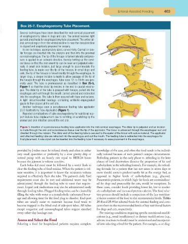
Another technique uses a percutaneous feeding tube applicator
(ELD Gastrostomy Tube Applicator) (Figure 1).
Reported complications of tube esophagostomy for nutritional sup-
port include tube displacement due to vomiting or scratching by the
animal and skin infection around the exit site.
Figure 1. Insertion of a percutaneous feeding tube applicator into the mid-cervical esophagus. The distal tip is palpated and an incision
is made through the skin and subcutaneous tissue over the tip of the applicator. The trocar is advanced through the esophageal wall and
directed through the incision. The distal end of the feeding tube is secured to the eyelet of the trocar with suture material. The applicator
and attached feeding tube are retracted into the esophagus and out the mouth. The feeding tube is redirected into the esophagus for
final placement. A wire stylet can be inserted into the feeding tube if necessary to ease placement in the esophagus.
provided by J-tubes must be infused slowly and often in either knowledge of the case, and often the food needs to be individ-
very small quantities or preferably by a slow gravity drip or ually tailored because of each patient’s unique circumstances.
enteral pump with an hourly rate equal to RER/24 hours Refeeding patients in the early phase vs. refeeding in the later
because the jejunum is volume sensitive. phases of food deprivation dictates the proportion of fat and
Each bolus-fed meal must be followed by a water flush to carbohydrate in the refeeding formula. For example, the refeed-
clear the feeding tube of food residue. When the patient is vol- ing formula for a patient that has not eaten in seven days or
ume sensitive, it is important to know the minimum volume more should contain predominantly fat as the energy fuel, as
required to effectively flush the tube. The patient’s daily fluid opposed to higher levels of carbohydrate (e.g., glucose).
requirement must also be met and additional water may be Pancreatitis patients, in which high-fat foods are contraindicat-
administered through the feeding tube to meet that require- ed for dogs and presumably for cats, would be exceptions. In
ment. Liquid oral medications may also be administered easily these cases, consider foods providing lower fat, low to moder-
through feeding tubes. Plugged feeding tubes can be cleared by ate carbohydrate and increased protein calories.The food selec-
filling the tube with water or a nonalcoholic carbonated bever- tion process should include a comparison of the key nutrition-
age and allowing time for the food plug to dissolve. End-port al factor content of the food to the recommended levels. Tables
tubes are usually easier to maintain because food tends to 25-8 and 25-9 list selected foods for assisted feeding and com-
become trapped in the blind end of side-port tubes. All tubes pares them to the recommended levels of key nutritional factors
except orogastric and nasoesophageal tubes require standard for dogs and cats, respectively.
every-other-day bandage care. Pre-existing conditions requiring specific nutritional modifi-
cations (e.g., renal insufficiency) or dietary modifications (e.g.,
Assess and Select the Food adverse reactions to foods) must be understood and incorporat-
Selecting a food for hospitalized patients requires complete ed into selecting a food for the patient. For example, a cat diag-