
Page 452 - Small Animal Clinical Nutrition 5th Edition
P. 452
466 Small Animal Clinical Nutrition
VetBooks.ir Box 25-9. Percutaneous Endoscopic Gastrostomy Tubes.
Percutaneous endoscopic gastrostomy (PEG) tubes are inserted with days. Firm adhesions between the gastric serosa and the peritoneum
the aid of general anesthesia. The patient is placed in right lateral have been reported to form within 36 to 48 hours of PEG tube place-
recumbency and an area of the left flank extending 4 to 6 inches ment in healthy dogs but do not reliably form in healthy cats.
caudal to the last rib is surgically prepared. Figures 1 to 7 describe Adhesion formation may also be variable in undernourished animals.
tube placement technique in detail. Landmarks for feeding tube The stomach should be empty when the tube is removed.
placement are usually 1 to 2 cm caudal to the last rib and one-third Sedation or anesthesia is not generally required for tube extraction.
the distance from the ventral border of the epaxial musculature to Tubes are removed by exerting firm traction on the tube, while simul-
the ventral midline. Commercial PEG catheter assembly kits, ranging taneously applying counter-pressure around the exit site (Figure 8).
in size from 16 to 28 Fr., are now available for small animal patients An alternative method of removal, suitable for dogs weighing more
and provide cost-effective, convenient materials for PEG tube place- than 10 kg, is to cut the catheter off flush with the skin, leaving the
ment (Figure 4). catheter tip to be passed in the feces. The resulting gastrocutaneous
Following insertion, the tube is usually incorporated into a light fistula usually heals rapidly.
bandage, with the free end brought to a convenient position for feed- Complications of PEG tube placement include vomiting, peristom-
ing. PEG tubes should be left in place for a minimum of five to seven al skin infection, cellulitis and pressure necrosis at the tube exit site.
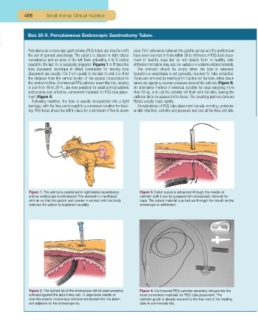
Figure 1. The animal is positioned in right lateral recumbency Figure 3. Nylon suture is advanced through the needle or
and an endoscope is introduced. The stomach is insufflated catheter until it can be grasped with endoscopic retrieval for-
with air so that the gastric wall comes in contact with the body ceps. The suture material is pulled out through the mouth as the
wall and the spleen is displaced caudally. endoscope is withdrawn.
Figure 2. The lighted tip of the endoscope will be seen pressing Figure 4. Commercial PEG catheter assembly kits provide the
outward against the abdominal wall. A large-bore needle or most convenient materials for PEG tube placement. The
over-the-needle intravenous catheter is inserted into the stom- catheter guide is already secured to the free end of the feeding
ach adjacent to the endoscope tip. tube in commercial kits.