
Page 448 - Small Animal Clinical Nutrition 5th Edition
P. 448
462 Small Animal Clinical Nutrition
VetBooks.ir In some instances, a pharyngostomy tube is used to bypass the
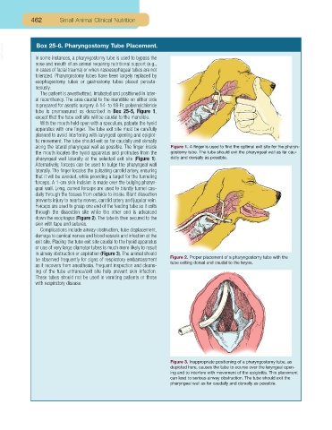
Box 25-6. Pharyngostomy Tube Placement.
nose and mouth of an animal requiring nutritional support (e.g.,
in cases of facial trauma) or when nasoesophageal tubes are not
tolerated. Pharyngostomy tubes have been largely replaced by
esophagostomy tubes or gastrostomy tubes placed percuta-
neously.
The patient is anesthetized, intubated and positioned in later-
al recumbency. The area caudal to the mandible on either side
is prepared for aseptic surgery. A 14- to 18-Fr. polyvinylchloride
tube is premeasured as described in Box 25-5, Figure 1,
except that the tube exit site will be caudal to the mandible.
With the mouth held open with a speculum, palpate the hyoid
apparatus with one finger. The tube exit site must be carefully
planned to avoid interfering with laryngeal opening and epiglot-
tic movement. The tube should exit as far caudally and dorsally
along the lateral pharyngeal wall as possible. The finger inside Figure 1. A finger is used to find the optimal exit site for the pharyn-
the mouth locates the hyoid apparatus and protrudes from the gostomy tube. The tube should exit the pharyngeal wall as far cau-
pharyngeal wall laterally at the selected exit site (Figure 1). dally and dorsally as possible.
Alternatively, forceps can be used to bulge the pharyngeal wall
laterally. The finger locates the pulsating carotid artery, ensuring
that it will be avoided, while providing a target for the tunneling
forceps. A 1-cm skin incision is made over the bulging pharyn-
geal wall. Long, curved forceps are used to bluntly tunnel cau-
dally through the tissues from outside to inside. Blunt dissection
prevents injury to nearby nerves, carotid artery and jugular vein.
Forceps are used to grasp one end of the feeding tube so it exits
through the dissection site while the other end is advanced
down the esophagus (Figure 2). The tube is then secured to the
skin with tape and sutures.
Complications include airway obstruction, tube displacement,
damage to cervical nerves and blood vessels and infection at the
exit site. Placing the tube exit site caudal to the hyoid apparatus
or use of very large diameter tubes is much more likely to result
in airway obstruction or aspiration (Figure 3). The animal should
be observed frequently for signs of respiratory embarrassment Figure 2. Proper placement of a pharyngostomy tube with the
as it recovers from anesthesia. Frequent inspection and cleans- tube exiting dorsal and caudal to the larynx.
ing of the tube entrance/exit site help prevent skin infection.
These tubes should not be used in vomiting patients or those
with respiratory disease.
Figure 3. Inappropriate positioning of a pharyngostomy tube, as
depicted here, causes the tube to course over the laryngeal open-
ing and to interfere with movement of the epiglottis. This placement
can lead to serious airway obstruction. The tube should exit the
pharyngeal wall as far caudally and dorsally as possible.