
Page 96 - Clinical Pearls in Cardiology
P. 96
84 Clinical Pearls in Cardiology
Contd...
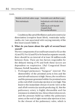
Mobile and thick valve cusps Immobile and calcified cusps
Thin individuals Individuals with thick chest
walls
Individuals with barrel-
shaped chest
Pericardial effusion
Conditions like atrial fibrillation and atrioventricular
dissociation (complete heart block, ventricular tachy-
cardia, etc.) are associated with varying intensity of the
first heart sound (Table 2).
3. What do you know about the split of second heart
sound?
The two components of second heart sound or S2 are the
A2 and P2. For A2 and P2 to be heard as separate sounds,
there should be an interval of at least 30 milliseconds
between them. There are two factors responsible for
the delayed timing of P2 and both these factors are
dependent on respiration. (Ref: Congenital Heart
Disease, Perloff. Saunders; 2012)
1. Prolonged pulmonary “hangout” interval: The
distensibility of the proximal aorta is less and the
arterial wall resistance is high. Hence, the recoil force
and the pressure generated within the proximal aorta
at the end of left ventricular ejection is very high. This
leads to the immediate closure of aortic valve at the
end of left ventricular systole producing A2. But the
pulmonary artery is highly distensible and the
resistance is relatively less. Hence there is less recoil,
and the pressure build up within the pulmonary
artery at the end of right ventricular ejection is less.
So the closure of the pulmonary valve, which is