
Page 97 - Essential Haematology
P. 97
Chapter 6 Haemolytic anaemias / 83
complement, and are therefore taken up by RE Laboratory fi ndings
macrophages which have receptors for the Ig Fc Th e haematological and biochemical fi ndings are
fragment. Part of the coated membrane is lost so typical of an extravascular haemolytic anaemia with
the cell becomes progressively more spherical to spherocytosis prominent in the peripheral blood
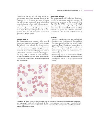
maintain the same volume and is ultimately prema- (Fig. 6.9 a). The DAT is positive as a result of IgG,
turely destroyed, predominantly in the spleen. IgG and complement or IgA on the cells and, in
When the cells are coated with IgG and comple- some cases, the autoantibody shows specifi city
ment (C3d, the degraded fragment of C3) or com- within the Rh system. The antibodies both on the
plement alone, red cell destruction occurs more cell surface and free in serum are best detected at
generally in the RE system. 37 ° C.
Treatment
Clinical f eatures 1 Remove the underlying cause (e.g. methyldopa).
The disease may occur at any age, in either sex, and 2 Corticosteroids. Prednisolone is the usual fi rst -
presents as a haemolytic anaemia of varying severity. line treatment; 60 mg/day is a typical starting
The spleen is often enlarged. The disease tends to dose in adults and should then be tapered down.
remit and relapse. It may occur alone or in associa- Those with predominantly IgG on red cells do
tion with other diseases (Table 6.5 ). When associ- best whereas those with complement often
ated with idiopathic thrombocytopenic purpura respond poorly, both to corticosteroids and
(ITP), which is a similar condition aff ecting plate- splenectomy.
lets (see p. 334) , it is known as Evans ’ syndrome. 3 Splenectomy may be of value in those who fail
When secondary to systemic lupus erythematosus to respond well or fail to maintain a satisfactory
the cells typically are coated with immunoglobulin haemoglobin level on an acceptably small steroid
and complement. dosage.
(a) (b)
Figure 6.9 (a) Blood fi lm in warm autoimmune haemolytic anaemia. Numerous microspherocytes are present
and larger polychromatic cells (reticulocytes). (b) Blood fi lm in cold autoimmune haemolytic anaemia. Marked
red cell agglutination is present in fi lms made at room temperature. The background is caused by the raised
plasma protein concentration.