
Page 92 - Essential Haematology
P. 92
78 / Chapter 6 Haemolytic anaemias
Pathogenesis Clinical f eatures
HS is usually caused by defects in the proteins The inheritance is autosomal dominant with varia-
involved in the vertical interactions between the ble expression; rarely it may be autosomal recessive.
membrane skeleton and the lipid bilayer of the red The anaemia can present at any age from infancy to
cell (Table 6.3 ; see Fig. 2.12 ). The loss of membrane old age. Jaundice is typically fluctuating and is par-
may be caused by the release of parts of the lipid ticularly marked if the haemolytic anaemia is associ-
’
bilayer that are not supported by the skeleton. In ated with Gilbert s disease (a defect of hepatic
HS, the marrow produces red cells of normal bicon- conjugation of bilirubin); splenomegaly occurs in
cave shape but these lose membrane and become most patients. Pigment gallstones are frequent (Fig.
increasingly spherical (loss of surface area relative to 6.2 ); aplastic crises, usually precipitated by parvovi-
volume) as they circulate through the spleen and rus infection, may cause a sudden increase in sever-
the rest of the RE system. Ultimately, the sphero- ity of anaemia (see Fig. 22.5 ).
cytes are unable to pass through the splenic micro-
circulation where they die prematurely.
Haematological fi ndings
Anaemia is usual but not invariable; its severity
Table 6.3 Molecular basis of hereditary tends to be similar in members of the same family.
spherocytosis and elliptocytosis. Reticulocytes are usually 5 – 20%. The blood fi lm
shows microspherocytes (Fig. 6.4 a) that are densely
Hereditary spherocytosis staining with smaller diameters than normal red
Ankyrin defi ciency or abnormalities cells.
α - or β - spectrin defi ciency or abnormalities
Band 3 abnormalities
Pallidin (protein 4.2) abnormalities Investigation and t reatment
A rapid fl uorescent flow analysis of eosin - maleimide
Hereditary elliptocytosis
bound to red cells is used as a test for HS and
α - or β - spectrin mutants leading to defective
membrane band 3 protein deficiency (Fig. 6.5 ).
spectrin dimer formation
This has replaced the classic osmotic fragility test
α - or β - spectrin mutants leading to defective
spectrin – ankyrin associations which showed the HS red cells to be excessively
fragile in dilute saline solution. Th e identifi cation
Protein 4.1 defi ciency or abnormality
of the exact molecular defect is not needed for man-
South - East Asian ovalocytosis (band 3 deletion)
agement. The direct antiglobulin (Coombs) test is
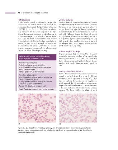
(a) (b)
Figure 6.4 (a) Blood fi lm in hereditary spherocytosis. The spherocytes are deeply staining and of small
diameter. Larger polychromatic cells are reticulocytes (confi rmed by supravital staining). (b) Blood fi lm in
hereditary elliptocytosis.