
Page 95 - Essential Haematology
P. 95
Chapter 6 Haemolytic anaemias / 81
young red cells, red cell enzyme assay may give a
Table 6.4 Agents that may cause haemolytic
anaemia in glucose - 6 - phosphate ‘ false ’ normal level in the phase of acute haemolysis
dehydrogenase (G6PD) defi ciency. with a reticulocyte response. Subsequent assay
after the acute phase reveals the low G6PD level
Infections and other acute illnesses (e.g. diabetic when the red cell population is of normal age
ketoacidosis) distribution.
Drugs
Treatment
Antimalarials (e.g. primaquine, pamaquine, Th e offending drug is stopped, any underlying
®
®
chloroquine, Fansidar , Maloprim ) infection is treated, a high urine output is main-
Sulphonamides and sulphones (e.g. co - tained and blood transfusion undertaken where
trimoxazole, sulfanilamide, dapsone, necessary for severe anaemia. G6PD - defi cient
®
Salazopyrin ) babies are prone to neonatal jaundice and in severe
cases phototherapy and exchange transfusion may
Other antibacterial agents (e.g. nitrofurans,
be needed. The jaundice is usually not caused by
chloramphenicol)
excess haemolysis but by deficiency of G6PD aff ect-
Analgesics (e.g. aspirin), moderate doses are ing neonatal liver function.
safe
Antihelminths (e.g. β - naphthol, stibophen)
Glutathione d eficiency and o ther
Miscellaneous (e.g. vitamin K analogues, s yndromes
naphthalene (mothballs), probenecid)
Other defects in the pentose phosphate pathway
Fava beans (possibly other vegetables)
leading to similar syndromes to G6PD defi ciency
have been described – particularly glutathione
NB. Many common drugs have been reported to
precipitate haemolysis in G6PD defi ciency in some defi ciency.
patients (e.g. aspirin, quinine and penicillin) but not at
conventional dosage.
Glycolytic (Embden – Meyerhof)
p athway d efects
These are all uncommon and lead to a congenital
non - spherocytic haemolytic anaemia. In some there
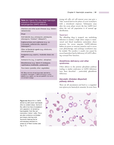
Figure 6.8 Blood fi lm in G6PD
defi ciency with acute haemolysis
after an oxidant stress. Some of
the cells show loss of cytoplasm
with separation of remaining
haemoglobin from the cell
membrane ( ‘ blister ’ cells). There
are also numerous contracted
and deeply staining cells.
Supravital staining (as for
reticulocytes) showed the
presence of Heinz bodies (see
Fig. 2.17 ).