Page 91 - Essential Haematology
P. 91

Chapter 6  Haemolytic anaemias  /  77


                                                                also formed from the process of intravascular

                            Table 6.2   Causes of intravascular
                        haemolysis.                             haemolysis.
                                                                    The main laboratory features of intravascular

                                                                haemolysis are:
                            Mismatched blood transfusion (usually ABO)
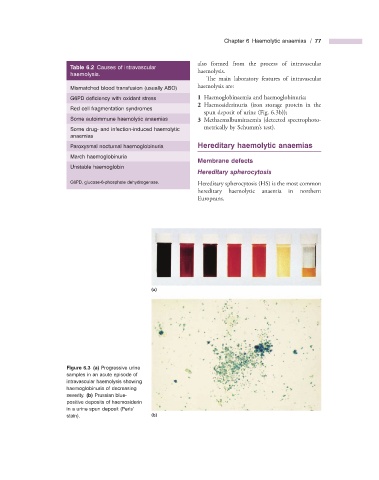
                            G6PD defi ciency with oxidant stress        1      Haemoglobinaemia and haemoglobinuria;
                                                                   2      Haemosiderinuria (iron storage protein in the
                            Red cell fragmentation syndromes
                                                                  spun deposit of urine (Fig.  6.3 b));
                            Some autoimmune haemolytic anaemias        3      Methaemalbuminaemia (detected spectrophoto-
                            Some drug -  and infection - induced haemolytic   metrically by Schumm ’ s test).
                        anaemias
                            Paroxysmal nocturnal haemoglobinuria         Hereditary  h aemolytic  a naemias
                            March haemoglobinuria
                                                                    Membrane  d efects
                            Unstable haemoglobin
                                                                    Hereditary  s pherocytosis
                              G6PD, glucose - 6 - phosphate dehydrogenase.       Hereditary spherocytosis (HS) is the most common
                                                                hereditary haemolytic anaemia in northern
                                                                Europeans.


                                                  (a)


                                Figure 6.3   (a)  Progressive urine

                      samples in an acute episode of
                      intravascular haemolysis showing
                      haemoglobinuria of decreasing
                      severity.  (b)  Prussian blue -
                        positive deposits of haemosiderin
                      in a urine spun deposit (Perls ’
                      stain).                     (b)
   86   87   88   89   90   91   92   93   94   95   96