
Page 90 - Essential Haematology
P. 90
76 / Chapter 6 Haemolytic anaemias
on standing because of excess urobilinogen. Pigment (b) Bone marrow erythroid hyperplasia; the
(bilirubin) gallstones may complicate the condition normal marrow myeloid : erythroid ratio of
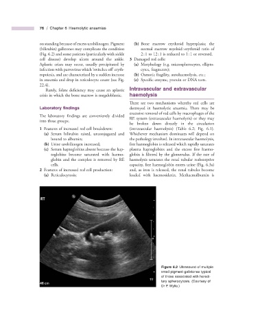
(Fig. 6.2 ) and some patients (particularly with sickle 2 : 1 to 12 : 1 is reduced to 1 : 1 or reversed.
cell disease) develop ulcers around the ankle. 3 Damaged red cells:
Aplastic crises may occur, usually precipitated by (a) Morphology (e.g. microspherocytes, ellipto-
‘
infection with parvovirus which switches off ’ eryth- cytes, fragments);
ropoiesis, and are characterized by a sudden increase (b) Osmotic fragility, autohaemolysis, etc.;
in anaemia and drop in reticulocyte count (see Fig. (c) Specific enzyme, protein or DNA tests.
22.4 ).
Rarely, folate deficiency may cause an aplastic Intravascular and e xtravascular
crisis in which the bone marrow is megaloblastic. h aemolysis
There are two mechanisms whereby red cells are
Laboratory fi ndings destroyed in haemolytic anaemia. There may be
excessive removal of red cells by macrophages of the
The laboratory findings are conveniently divided
RE system (extravascular haemolysis) or they may
into three groups.
be broken down directly in the circulation
1 Features of increased red cell breakdown: (intravascular haemolysis) (Table 6.2 ; Fig. 6.1 ).
(a) Serum bilirubin raised, unconjugated and Whichever mechanism dominates will depend on
bound to albumin; the pathology involved. In intravascular haemolysis,
(b) Urine urobilinogen increased; free haemoglobin is released which rapidly saturates
(c) Serum haptoglobins absent because the hap- plasma haptoglobins and the excess free haemo-
toglobins become saturated with haemo- globin is filtered by the glomerulus. If the rate of
globin and the complex is removed by RE haemolysis saturates the renal tubular reabsorptive
cells. capacity, free haemoglobin enters urine (Fig. 6.3 a)
2 Features of increased red cell production: and, as iron is released, the renal tubules become
(a) Reticulocytosis; loaded with haemosiderin. Methaemalbumin is
Figure 6.2 Ultrasound of multiple
small pigment gallstones typical
of those associated with heredi-
tary spherocytosis. (Courtesy of
Dr P. Wylie.)