
Page 623 - Basic _ Clinical Pharmacology ( PDFDrive )
P. 623
CHAPTER 34 Drugs Used in Disorders of Coagulation 609
Wall defect
EC
C vWF
GP Ia GP Ib
ADP PGI
TXA 2 2
5-HT –
GP Intrinsic Extrinsic
IIb/IIIa
Platelets Xa
Degranulation
+ GP + +
+ IIb/IIIa Activation
+
GP +
IIb/IIIa
Fibrin
Thrombin Prothrombin
+
Fibrinogen
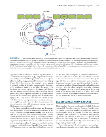
FIGURE 34–1 Thrombus formation at the site of the damaged vascular wall (EC, endothelial cell) and the role of platelets and clotting fac-
tors. Platelet membrane receptors include the glycoprotein (GP) Ia receptor, binding to collagen (C); GP Ib receptor, binding von Willebrand fac-
tor (vWF); and GP IIb/IIIa, which binds fibrinogen and other macromolecules. Antiplatelet prostacyclin (PGI 2 ) is released from the endothelium.
Aggregating substances released from the degranulating platelet include adenosine diphosphate (ADP), thromboxane A 2 (TXA 2 ), and serotonin
(5-HT). Production of factor Xa by intrinsic and extrinsic pathways is detailed in Figure 34–2. (Redrawn and reproduced, with permission, from Simoons ML,
Decker JW: New directions in anticoagulant and antiplatelet treatment. [Editorial.] Br Heart J 1995;74:337.)
aggregation and vasoconstriction. Activation of platelets results in but the most feared consequence is pulmonary embolism (PE).
a conformational change in the α β integrin (IIb/IIIa) recep- This occurs when part or all of the clot breaks off from its location
IIb III
tor, enabling it to bind fibrinogen, which cross-links adjacent in the deep venous system and travels as an embolus through the
platelets, resulting in aggregation and formation of a platelet plug right side of the heart and into the pulmonary arterial circulation.
(Figure 34–1). Simultaneously, the coagulation system cascade Occlusion of a large pulmonary artery by an embolic clot can pre-
is activated, resulting in thrombin generation and a fibrin clot, cipitate acute right heart failure and sudden death. In addition lung
which stabilizes the platelet plug (see below). Knowledge of the ischemia or infarction will occur distal to the occluded pulmonary
hemostatic mechanism is important for diagnosis of bleeding arterial segment. Such emboli usually arise from the deep venous
disorders. Patients with defects in the formation of the primary system of the proximal lower extremities or pelvis. Although all
platelet plug (defects in primary hemostasis, eg, platelet function thrombi are mixed, the platelet nidus dominates the arterial throm-
defects, von Willebrand disease) typically bleed from surface sites bus and the fibrin tail dominates the venous thrombus.
(gingiva, skin, heavy menses) with injury. In contrast, patients
with defects in the clotting mechanism (secondary hemostasis,
eg, hemophilia A) tend to bleed into deep tissues (joints, muscle, BLOOD COAGULATION CASCADE
retroperitoneum), often with no apparent inciting event, and
bleeding may recur unpredictably. Blood coagulates due to the transformation of soluble fibrinogen
The platelet is central to normal hemostasis and thromboem- into insoluble fibrin by the enzyme thrombin. Several circulat-
bolic disease, and is the target of many therapies discussed in this ing proteins interact in a cascading series of limited proteolytic
chapter. Platelet-rich thrombi (white thrombi) form in the high reactions (Figure 34–2). At each step, a clotting factor zymogen
flow rate and high shear force environment of arteries. Occlusive undergoes limited proteolysis and becomes an active protease
arterial thrombi cause serious disease by producing downstream (eg, factor VII is converted to factor VIIa). Each protease factor
ischemia of extremities or vital organs, and they can result in limb activates the next clotting factor in the sequence, culminating in
amputation or organ failure. Venous clots tend to be more fibrin- the formation of thrombin (factor IIa). Several of these factors are
rich, contain large numbers of trapped red blood cells, and are targets for drug therapy (Table 34–1).
recognized pathologically as red thrombi. Deep venous thrombi Thrombin has a central role in hemostasis and has many func-
(DVT) can cause severe swelling and pain of the affected extremity, tions. In clotting, thrombin proteolytically cleaves small peptides