
Page 625 - Basic _ Clinical Pharmacology ( PDFDrive )
P. 625
CHAPTER 34 Drugs Used in Disorders of Coagulation 611
VIII and IX. (This provides an explanation for why patients with
deficiency of factor VIII or IX—hemophilia A and hemophilia B, Plasminogen
respectively—have a severe bleeding disorder.) Activation Inhibition
It is also important to note that the coagulation mechanism Various stimuli
in vivo does not occur in solution, but is localized to activated
cell surfaces expressing anionic phospholipids such as phosphati- +
2+
dylserine, and is mediated by Ca bridging between the anionic Blood Blood + − Antiactivators
phospholipids and γ-carboxyglutamic acid residues of the clotting proactivator activator
factors. This is the basis for using calcium chelators such as eth- t-PA, urokinase + − Aminocaproic
ylenediamine tetraacetic acid (EDTA) or citrate to prevent blood acid
from clotting in a test tube. Streptokinase
Antithrombin (AT) is an endogenous anticoagulant and a Activator +
member of the serine protease inhibitor (serpin) family; it inacti-
vates the serine proteases IIa, IXa, Xa, XIa, and XIIa. The endog- Proactivator Plasmin
enous anticoagulants protein C and protein S attenuate the blood Anistreplase
clotting cascade by proteolysis of the two cofactors Va and VIIIa. + +
From an evolutionary perspective, it is of interest that factors V
and VIII have an identical overall domain structure and consider- Fibrinogen Thrombin Fibrin
degradation
able homology, consistent with a common ancestor gene; likewise degradation Fibrinogen Fibrin clot products,
products
the serine proteases are descendants of a trypsin-like common Factor XIII D-dimer
ancestor. Thus, the TF-VIIa initiating complex, serine proteases,
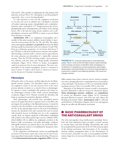
and cofactors each have their own lineage-specific attenuation FIGURE 34–3 Schematic representation of the fibrinolytic
mechanism (Figure 34–2). Defects in natural anticoagulants system. Plasmin is the active fibrinolytic enzyme. Several clinically
result in an increased risk of venous thrombosis. The most com- useful activators are shown on the left in bold. Anistreplase is a
mon defect in the natural anticoagulant system is a mutation in combination of streptokinase and the proactivator plasminogen.
factor V (factor V Leiden), which results in resistance to inactiva- Aminocaproic acid (right) inhibits the activation of plasminogen to
tion by the protein C/protein S mechanism. plasmin and is useful in some bleeding disorders. t-PA, tissue plas-
minogen activator.
Fibrinolysis
follow massive tissue injury, advanced cancers, obstetric emergen-
Fibrinolysis refers to the process of fibrin digestion by the fibrin- cies such as abruptio placentae or retained products of conception,
specific protease, plasmin. The fibrinolytic system is similar to or bacterial sepsis. The treatment of DIC is to control the underly-
the coagulation system in that the precursor form of the serine ing disease process; if this is not possible, DIC is often fatal.
protease plasmin circulates in an inactive form as plasminogen. Regulation of the fibrinolytic system is useful in therapeutics.
In response to injury, endothelial cells synthesize and release tis- Increased fibrinolysis is effective therapy for thrombotic disease.
sue plasminogen activator (t-PA), which converts plasminogen Tissue plasminogen activator, urokinase, and streptokinase
to plasmin (Figure 34–3). Plasmin remodels the thrombus and all activate the fibrinolytic system (Figure 34–3). Conversely,
limits its extension by proteolytic digestion of fibrin. decreased fibrinolysis protects clots from lysis and reduces the
Both plasminogen and plasmin have specialized protein bleeding of hemostatic failure. Aminocaproic acid is a clinically
domains (kringles) that bind to exposed lysines on the fibrin clot useful inhibitor of fibrinolysis. Heparin and the oral anticoagulant
and impart clot specificity to the fibrinolytic process. It should be drugs do not affect the fibrinolytic mechanism.
noted that this clot specificity is only observed at physiologic levels
of t-PA. At the pharmacologic levels of t-PA used in thrombolytic
therapy, clot specificity is lost and a systemic lytic state is created,
with attendant increase in bleeding risk. As in the coagulation ■ BASIC PHARMACOLOGY OF
cascade, there are negative regulators of fibrinolysis: endothelial THE ANTICOAGULANT DRUGS
cells synthesize and release plasminogen activator inhibitor (PAI),
antiplasmin circulates in the The ideal anticoagulant drug would prevent pathologic throm-
which inhibits t-PA; in addition α 2
blood at high concentrations and under physiologic conditions bosis and limit reperfusion injury yet allow a normal response
will rapidly inactivate any plasmin that is not clot-bound. How- to vascular injury and limit bleeding. Theoretically this could
ever, this regulatory system is overwhelmed by therapeutic doses be accomplished by preservation of the TF-VIIa initiation phase
of plasminogen activators. of the clotting mechanism with attenuation of the secondary
If the coagulation and fibrinolytic systems are pathologically intrinsic pathway propagation phase of clot development. At
activated, the hemostatic system may careen out of control, lead- this time such a drug does not exist; all anticoagulants and fibri-
ing to generalized intravascular clotting and bleeding. This process nolytic drugs have an increased bleeding risk as their principle
is called disseminated intravascular coagulation (DIC) and may toxicity.