
Page 129 - Atlas of Histology with Functional Correlations
P. 129
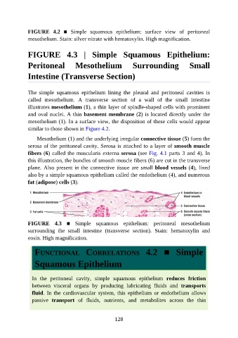
FIGURE  4.2  ■  Simple  squamous  epithelium:  surface  view  of  peritoneal

               mesothelium. Stain: silver nitrate with hematoxylin. High magnification.


               FIGURE  4.3  |  Simple  Squamous  Epithelium:


               Peritoneal              Mesothelium                 Surrounding                  Small

               Intestine (Transverse Section)


               The  simple  squamous  epithelium  lining  the  pleural  and  peritoneal  cavities  is
               called  mesothelium.  A  transverse  section  of  a  wall  of  the  small  intestine

               illustrates mesothelium (1), a thin layer of spindle-shaped cells with prominent
               and oval nuclei. A thin basement membrane (2) is located directly under the
               mesothelium (1). In a surface view, the disposition of these cells would appear
               similar to those shown in Figure 4.2.

                   Mesothelium (1) and the underlying irregular connective tissue (5) form the

               serosa of the peritoneal cavity. Serosa is attached to a layer of smooth muscle
               fibers (6) called the muscularis externa serosa (see Fig. 4.1 parts 3 and 4). In
               this illustration, the bundles of smooth muscle fibers (6) are cut in the transverse

               plane. Also present in the connective tissue are small blood vessels (4), lined
               also by a simple squamous epithelium called the endothelium (4), and numerous
               fat (adipose) cells (3).


               FIGURE  4.3  ■  Simple  squamous  epithelium:  peritoneal  mesothelium
               surrounding  the  small  intestine  (transverse  section).  Stain:  hematoxylin  and
               eosin. High magnification.


                  FUNCTIONAL  CORRELATIONS  4.2  ■  Simple

                  Squamous Epithelium


                 In  the  peritoneal  cavity,  simple  squamous  epithelium  reduces  friction
                 between  visceral  organs  by  producing  lubricating  fluids  and  transports
                 fluid.  In  the  cardiovascular  system,  this  epithelium  or  endothelium  allows

                 passive  transport  of  fluids,  nutrients,  and  metabolites  across  the  thin


                                                          128