
Page 134 - Atlas of Histology with Functional Correlations
P. 134
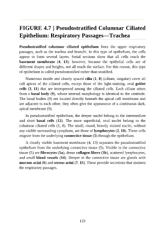
FIGURE 4.7 | Pseudostratified Columnar Ciliated

               Epithelium: Respiratory Passages—Trachea


               Pseudostratified  columnar  ciliated  epithelium  lines  the  upper  respiratory

               passages, such as the trachea and bronchi. In this type of epithelium, the cells
               appear  to  form  several  layers.  Serial  sections  show  that  all  cells  reach  the
               basement  membrane  (4,  13);  however,  because  the  epithelial  cells  are  of
               different shapes and heights, not all reach the surface. For this reason, this type

               of epithelium is called pseudostratified rather than stratified.

                   Numerous motile and closely spaced cilia (1, 8) (cilium, singular) cover all
               cell  apices  of  the  ciliated  cells,  except  those  of  the  light-staining,  oval  goblet
               cells (3, 11)  that  are  interspersed  among  the  ciliated  cells.  Each  cilium  arises

               from a basal body (9), whose internal morphology is identical to the centriole.
               The basal bodies (9) are located directly beneath the apical cell membrane and
               are adjacent to each other; they often give the appearance of a continuous dark,
               apical membrane (9).

                   In pseudostratified epithelium, the deeper nuclei belong to the intermediate

               and  short  basal  cells  (12).  The  more  superficial,  oval  nuclei  belong  to  the
               columnar ciliated cells (1, 8). The small, round, heavily stained nuclei, without
               any visible surrounding cytoplasm, are those of lymphocytes (2, 10). These cells
               migrate from the underlying connective tissue (5) through the epithelium.


                   A clearly visible basement membrane (4, 13) separates the pseudostratified
               epithelium from the underlying connective tissue (5). Visible in the connective
               tissue (5) are fibrocytes (5a), dense collagen fibers (5b), scattered lymphocytes,
               and small blood vessels (14). Deeper in the connective tissue are glands with
               mucous acini (6) and serous acini (7, 15). These provide secretions that moisten

               the respiratory passages.


                                                          133