
Page 136 - Atlas of Histology with Functional Correlations
P. 136
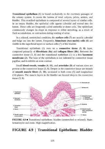
Transitional epithelium (1) is found exclusively in the excretory passages of

               the  urinary  system.  It  covers  the  lumina  of  renal  calyces,  pelvis,  ureters,  and
               bladder. This stratified epithelium is composed of several layers of similar cells.
               In  an  empty  bladder,  the  epithelial  cells  appear  cuboidal  and  extend  into  the
               lumen. These cells are frequently called umbrella or dome cells. The epithelium
               continuously  changes  its  shape  in  response  to  either  stretching,  as  a  result  of

               fluid accumulation, or contraction during voiding of urine.

                   In a relaxed, unstretched condition, the surface cells (7) are usually cuboidal
               and bulge out into the lumen. Frequently, binucleate (two nuclei) cells (6) are
               visible in the superficial layers or surface cells (7) of the bladder.


                   Transitional  epithelium  (1)  rests  on  a  connective  tissue  (3,  8)  layer,
               composed primarily of fibroblasts (8a) and collagen fibers (8b). Between the
               connective tissue (3, 8) and the transitional epithelium (1) is a thin basement
               membrane (2). The base of the epithelium is not indented by connective tissue
               papillae, and it exhibits an even contour.


                   Small blood vessels, venules (4, 11), and arterioles (9) of various sizes are
               present in the connective tissue (3, 8). Deeper in the connective tissue are strands
               of smooth muscle fibers (5, 10), sectioned in both cross (5) and longitudinal
               (10) planes. The muscle layers in the bladder are located deep to the connective

               tissue (3, 8).


               FIGURE 4.8 ■ Transitional epithelium: bladder (unstretched or relaxed). Stain:
               hematoxylin and eosin. High magnification.


               FIGURE  4.9  |  Transitional  Epithelium:  Bladder


                                                          135