
Page 139 - Atlas of Histology with Functional Correlations
P. 139
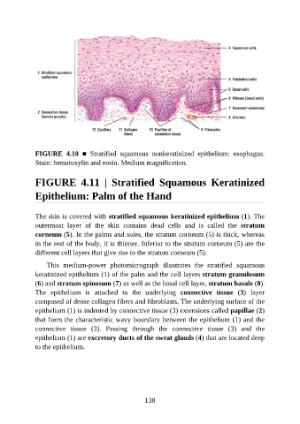
FIGURE  4.10  ■  Stratified  squamous  nonkeratinized  epithelium:  esophagus.
               Stain: hematoxylin and eosin. Medium magnification.


               FIGURE  4.11  |  Stratified  Squamous  Keratinized


               Epithelium: Palm of the Hand


               The skin is covered with stratified squamous keratinized epithelium (1). The
               outermost  layer  of  the  skin  contains  dead  cells  and  is  called  the  stratum

               corneum (5). In the palms and soles, the stratum corneum (5) is thick, whereas
               in the rest of the body, it is thinner. Inferior to the stratum corneum (5) are the
               different cell layers that give rise to the stratum corneum (5).

                   This  medium-power  photomicrograph  illustrates  the  stratified  squamous
               keratinized epithelium (1) of the palm and the cell layers stratum granulosum

               (6) and stratum spinosum (7) as well as the basal cell layer, stratum basale (8).
               The  epithelium  is  attached  to  the  underlying  connective  tissue  (3)  layer
               composed of dense collagen fibers and fibroblasts. The underlying surface of the
               epithelium (1) is indented by connective tissue (3) extensions called papillae (2)

               that form the characteristic wavy boundary between the epithelium (1) and the
               connective  tissue  (3).  Passing  through  the  connective  tissue  (3)  and  the
               epithelium (1) are excretory ducts of the sweat glands (4) that are located deep
               to the epithelium.


                                                          138