
Page 140 - Atlas of Histology with Functional Correlations
P. 140
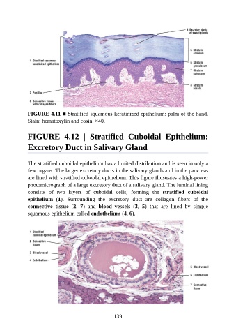
FIGURE 4.11 ■ Stratified squamous keratinized epithelium: palm of the hand.
               Stain: hematoxylin and eosin. ×40.


               FIGURE  4.12  |  Stratified  Cuboidal  Epithelium:

               Excretory Duct in Salivary Gland


               The stratified cuboidal epithelium has a limited distribution and is seen in only a
               few organs. The larger excretory ducts in the salivary glands and in the pancreas
               are lined with stratified cuboidal epithelium. This figure illustrates a high-power
               photomicrograph of a large excretory duct of a salivary gland. The luminal lining

               consists  of  two  layers  of  cuboidal  cells,  forming  the  stratified  cuboidal
               epithelium  (1).  Surrounding  the  excretory  duct  are  collagen  fibers  of  the
               connective  tissue  (2,  7)  and  blood  vessels  (3,  5)  that  are  lined  by  simple
               squamous epithelium called endothelium (4, 6).


                                                          139