
Page 131 - Atlas of Histology with Functional Correlations
P. 131
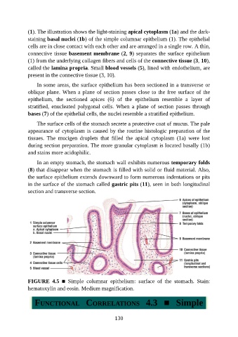
(1). The illustration shows the light-staining apical cytoplasm (1a) and the dark-

               staining basal nuclei (1b) of the simple columnar epithelium (1). The epithelial
               cells are in close contact with each other and are arranged in a single row. A thin,
               connective tissue basement membrane (2, 9) separates the surface epithelium
               (1) from the underlying collagen fibers and cells of the connective tissue (3, 10),
               called the lamina propria. Small blood vessels (5), lined with endothelium, are

               present in the connective tissue (3, 10).

                   In some areas, the surface epithelium has been sectioned in a transverse or
               oblique plane. When a plane of section passes close to the free surface of the
               epithelium,  the  sectioned  apices  (6)  of  the  epithelium  resemble  a  layer  of

               stratified,  enucleated  polygonal  cells.  When  a  plane  of  section  passes  through
               bases (7) of the epithelial cells, the nuclei resemble a stratified epithelium.

                   The surface cells of the stomach secrete a protective coat of mucus. The pale
               appearance of cytoplasm is caused by the routine histologic preparation of the
               tissues.  The  mucigen  droplets  that  filled  the  apical  cytoplasm  (1a)  were  lost

               during section preparation. The more granular cytoplasm is located basally (1b)
               and stains more acidophilic.

                   In an empty stomach, the stomach wall exhibits numerous temporary folds
               (8) that disappear when the stomach is filled with solid or fluid material. Also,

               the surface epithelium extends downward to form numerous indentations or pits
               in the surface of the stomach called gastric pits (11), seen in both longitudinal
               section and transverse section.


               FIGURE  4.5  ■  Simple  columnar  epithelium:  surface  of  the  stomach.  Stain:
               hematoxylin and eosin. Medium magnification.


                  FUNCTIONAL  CORRELATIONS  4.3  ■  Simple


                                                          130