
Page 173 - Atlas of Histology with Functional Correlations
P. 173
cells (1, 9) are usually ovoid, with a small, centrally placed nucleus and
cytoplasm filled with fine, closely packed granules that stain dense or deep red
with neutral red stain.
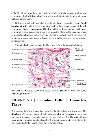
Different blood cells are also seen in the loose connective tissue. Small
lymphocytes (6) exhibit a dense-staining nucleus that occupies most of the cell
cytoplasm. Large lymphocytes (8) also exhibit a dense nucleus with more
cytoplasm. Loose connective tissue also contains blood cells eosinophils and
neutrophils and adipose cells. These are illustrated in greater detail in Figure 5.3,
in the loose connective tissue in Figure 5.5, and in the mesentery of an intestine
in Figure 5.13.
The faint background around the fibers and cells is the ground substance.
FIGURE 5.2 ■ Loose connective tissue (spread). Stained for cells and fibers.
High magnification.
FIGURE 5.3 | Individual Cells of Connective
Tissue
The main cells of the connective tissue are the fibroblasts and fibrocytes. The
fibroblast (1) is an elongated cell with cytoplasmic projections, an ovoid
nucleus with sparse chromatin, and one or two nucleoli. The fibrocyte (6) is a
more mature, smaller spindle-shaped cell without cytoplasmic projections; the
nucleus is similar but smaller than that in the fibroblast.
172