
Page 176 - Atlas of Histology with Functional Correlations
P. 176
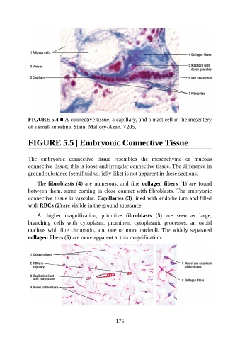
FIGURE 5.4 ■ A connective tissue, a capillary, and a mast cell in the mesentery

               of a small intestine. Stain: Mallory-Azan. ×205.


               FIGURE 5.5 | Embryonic Connective Tissue


               The  embryonic  connective  tissue  resembles  the  mesenchyme  or  mucous

               connective tissue; this is loose and irregular connective tissue. The difference in
               ground substance (semifluid vs. jelly-like) is not apparent in these sections.

                   The fibroblasts (4)  are  numerous,  and  fine  collagen fibers  (1)  are  found
               between  them,  some  coming  in  close  contact  with  fibroblasts.  The  embryonic

               connective tissue is vascular. Capillaries (3) lined with endothelium and filled
               with RBCs (2) are visible in the ground substance.

                   At  higher  magnification,  primitive  fibroblasts  (5)  are  seen  as  large,
               branching  cells  with  cytoplasm,  prominent  cytoplasmic  processes,  an  ovoid
               nucleus  with  fine  chromatin,  and  one  or  more  nucleoli.  The  widely  separated

               collagen fibers (6) are more apparent at this magnification.


                                                          175