
Page 179 - Atlas of Histology with Functional Correlations
P. 179
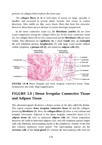
presence of collagen fibers replaces the loose type.

                   The  collagen  fibers  (2,  9)  in  both  types  of  tissues  are  large,  typically  in

               bundles,  and  sectioned  in  several  planes  because  they  course  in  various
               directions.  Also  visible  are  thin,  wavy  elastic  fibers  that  form  fine  networks.
               However, these fibers are not obvious in routine histologic preparations.

                   In  the  dense  connective  tissue  (1),  the  fibroblasts  (nuclei)  (3)  are  often
               found compressed among the collagen fibers (2). In the loose connective tissue

               (5), the collagen fibers (9) are less compressed and the fibroblasts (10) are more
               visible. Also illustrated are capillaries (4), a small venule (11), an eosinophil
               (6)  with  lobulated  nucleus,  lymphocytes  (7)  with  large  round  nuclei  without

               visible cytoplasm, a plasma cell (8), and numerous adipose cells (12).


               FIGURE  5.8  ■  Dense  irregular  and  loose  irregular  connective  tissue.  Stain:
               hematoxylin and eosin. High magnification.


               FIGURE 5.9 | Dense Irregular Connective Tissue

               and Adipose Tissue


               This photomicrograph illustrates a deeper section of the skin called the dermis.
               This  region  contains  dense  irregular  connective  tissue (1)  and  the  collagen-
               producing fibroblasts (3). Here, the collagen fibers (2) show a very random and
               irregular  orientation.  Adjacent  to  the  dense  irregular  connective  tissue  (1)  is

               adipose  tissue  (4)  with  its  numerous  adipose  cells  (5).  Tissue  preparation
               dissolves the lipids in individual adipose cells, and cell cytoplasm appears empty
               with only flattened, dense-staining nuclei in the peripheries. Dermis of the skin
               also  contains  numerous  sweat  glands.  The  light-staining  regions  are  the

               secretory cells of the sweat gland (7), whereas the dark-staining cells form the


                                                          178